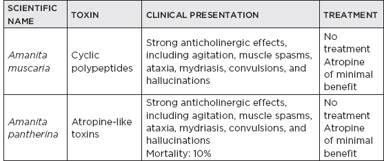
CHAPTER 15 Neurotoxicology and Nutritional Disorders I. Heavy Metals A. Arsenic 1. Pathophysiology: a primary source is pesticides; reacts with sulfhydryl groups of proteins and interferes with several steps of metabolism in the neuron, producing dying-back-type axonal degeneration, particularly in myelinated fibers 2. Clinical a. Axonal sensory neuropathy begins within 5 to 10 days. b. Acute gastrointestinal symptoms followed by painful paresthesia with progressive distal weakness c. Central nervous system (CNS) symptoms may develop rapidly in acute poisoning, with drowsiness and confusion progressing to stupor or delirium. d. Hyperkeratosis and sloughing of the skin on the palms and soles may occur several weeks after acute poisoning, followed by a chronic state of redness and swelling of the distal extremities. e. Nail changes (Mees’ lines) f. Chronic poisoning may develop aplastic anemia. 3. Diagnosis a. Acute intoxication: renal excretion greater than 0.1 mg arsenic in 24 hours b. Chronic intoxication: hair concentrations greater than 0.1 mg/100 g of hair 4. Treatment a. Acute oral ingestion i. Gastric lavage followed by instillation of 1% sodium thiosulfate ii. British anti-Lewisite (BAL) given parenterally in a 10% solution 5. Prognosis a. Once neuropathy occurs, treatment is usually ineffective. b. Mortality: greater than 50% to 75% in severe cases B. Gold 1. Pathophysiology: used in the treatment of inflammatory conditions 2. Clinical a. Chronic distal axonal sensory neuropathy more common than motor neuropathy b. Painful, involving palms or soles c. Myokymia d. Brachial plexopathy e. Acute inflammatory demyelinating polyradiculopathy 3. Pathology: loss of myelin as well as active axonal degeneration 4. Treatment: chelation therapy with BAL has been used but usually is not necessary. C. Mercury 1. Clinical a. Acute: salivation and severe gastrointestinal dysfunction followed by hallucinations and delirium b. Chronic i. Chronic axonal sensory neuropathy ii. Constriction of visual fields, ataxia, dysarthria, decreased hearing, tremor, and dementia iii. Parkinsonism iv. Children may have acrodynia. c. Minamata disease: methylmercury (MeHg) poisoning from ingestion of fish and shellfish contaminated by MeHg discharged in waste water; typical symptoms include sensory disturbances (glove and stocking type), ataxia, dysarthria, constriction of the visual field, auditory disturbances, and tremor 2. Treatment a. Chelating agents (e.g., D-penicillamine, BAL, ethylenediaminetetraacetic acid) D. Thallium 1. Pathophysiology: found in rat poison; thallium ions act interchangeably with potassium in respect to their transport by the Na/K ATPase system. 2. Clinical a. Hallmark: alopecia; sometimes with cranial nerve and autonomic involvement b. Acute i. Gastrointestinal symptoms within hours of ingestion ii. Moderate doses produce neuropathic symptoms in less than 48 hours consisting of pain and paresthesia, followed by ascending sensory loss and distal weakness. iii. May produce acute inflammatory demyelinating polyradiculopathy–like syndrome iv. Large doses (>2 g) produce cardiovascular shock, coma, and death within 24 hours. c. Chronic: chronic axonal sensorimotor neuropathy 3. Treatment a. Chelating agents i. Prussian blue (potassium ferric hexacyanoferrate) ii. BAL iii. Dithizone iv. Diethyldithiocarbamate b. If acute, can also perform gastric lavage E. Lead 1. Pathophysiology a. Diminishes cerebral glucose supplies b. Intoxication results in inhibition of myelin synthesis with demyelination. Lead has direct effects on porphyrin metabolism, by inhibiting gamma-aminolevulinic acid dehydrase. c. Adults i. Use of exterior paints and gasoline ii. More likely to present with neuropathy, predominantly, but not exclusively, the radial nerve d. Children i. Pica and eating lead-based paints ii. More likely to present with encephalopathy 2. Clinical a. Neuropathy i. Chronic axonal motor neuropathy ii. Classic neurologic presentation: wrist drop (Radial nerve palsy) iii. Typical clinical triad (A) Abdominal pain and constipation (B) Anemia (C) Neuropathy b. CNS toxicity i. Adult (A) Prodrome: progressive weakness and loss of weight (B) Ashen color of the face (C) Mild persistent headache (D) Fine tremor of the muscles of the eyes, tongue, and face (E) Progression into encephalopathic state (F) May have focal motor weakness ii. Children: prodrome usually nonspecific evolving into encephalopathy (50%) 3. Diagnosis a. Lead lines on gums (also known as Burtonian line)—stippled blue line seen in 50% to 70% of patients with chronic lead poisoning b. Serum: microcytic anemia and red blood cell basophilic stippling c. x-rays: may demonstrate lead lines of long bones 4. Treatment a. Chelating agents (e.g., BAL, ethylenediaminetetraacetic acid, penicillamine) 5. Prognosis a. Mild intoxication: usually complete recovery b. Severe encephalopathy: mortality high but lessened by the use of combined chelating agent therapy c. Residual neurologic sequelae: blindness or partial visual disturbances, persistent convulsions, personality changes, and mental retardation d. Prognosis worse in children than in adults F. Manganese 1. Pathology: diffuse injury to ganglion cells, primarily of globus pallidus 2. Clinical a. Extrapyramidal signs/symptoms, including dystonia, bradykinesia, tremor, and gait dysfunction b. Personality changes consisting of irritability, lack of sociability, uncontrollable laughter, tearfulness, and euphoria 3. Treatment: supportive therapy G. Iron 1. Acute iron toxicity a. Symptoms occur within 30 to 60 minutes. b. Initially, bloody vomiting followed by bloody diarrhea c. Severe cases: coma or convulsions d. Treatment i. Supportive care: induction of vomiting, gastric lavage, maintenance of adequate ventilation, correction of acidosis, and control of vital signs ii. Chelation: deferoxamine, 5 to 10 g e. Mortality: 45% 2. Iron deficiency can lead to restless leg syndrome. H. Tin: Triethyltin exposure acutely results in white-matter vacuolation; with chronic exposure, demyelination and gliosis are seen. II. Organic Solvents A. Methyl alcohol (e.g., methanol, wood alcohol) 1. Pathophysiology a. Component of antifreeze and alcoholic drinks b. Methanol itself is only mildly toxic, but its oxidation products (formaldehyde and formic acid) induce a severe acidosis. c. Methanol may cause bilateral hemorrhagic necrosis of the caudate, putamen, pons, optic nerves, cerebellum, and subcortical white matter. 2. Clinical a. Visual disturbance/ocular manifestations i. Amblyopia ii. Scotomas iii. Total blindness b. Extrapyramidal signs (bradykinesia, masked facies, tremor) 3. Treatment: three-part approach—ethanol, bicarbonate, dialysis (in severe cases) B. Ethylene glycol 1. Pathophysiology: used as antifreeze, tobacco moistener, and in paint; toxic dose is greater than 100 mL 2. Clinical a. Restless and agitation followed by somnolence, stupor, coma, and even convulsions b. Death due to cardiopulmonary failure c. Characteristic metabolic findings: metabolic acidosis with large anion gap, hypocalcemia, and calcium oxalate crystals in the urine 3. Treatment a. Supportive care b. Correct metabolic acidosis and hypocalcemia. c. Infuse ethanol at 5 to 10 g/hour. d. Dialysis may be necessary to remove ethylene glycol and to treat uremia. III. Gases A. Carbon monoxide poisoning 1. Pathophysiology a. Most common cause of death by poisoning in the United States b. Damages the brain by three mechanisms: i. Production of carboxyhemoglobin that causes hypoxemia ii. Decreased release of oxygen to tissues iii. Direct mitochondrial toxicity c. Pathology i. Bilateral necrotic lesions involving the globus pallidus ii. Hippocampal damage iii. Supratentorial demyelination iv. Cortical damage (watershed distribution) 2. Neuroimaging a. MRI: lesions usually appear hypointense on T1 and hyperintense on T2 involving globus pallidus. b. May also involve the thalamus, caudate, putamen, and cerebellum c. Differential diagnosis of bilateral basal ganglia lesions i. Carbon monoxide ii. Cyanide iii. Ethylene glycol iv. Methanol (more putamen) v. Aminoacidopathies vi. Infarction vii. Pantothenate-kinase-associated neurodegeneration (PKAN; formerly Hallervorden-Spatz disease) viii. Leigh disease ix. Wilson’s disease x. Mitochondrial disorders xi. Neoplasm xii. Multiple system atrophy 3. Clinical a. Hypoxia without cyanosis (cherry-red appearance) b. ± Myocardial infarction c. Retinal hemorrhages d. Neurologic: lethargy that progresses to coma followed by brainstem dysfunction and movement disorders 4. Treatment a. Oxygen 100% b. Hyperbaric chamber (if severe) 5. Prognosis: residual movement disorders are common. IV. Organophosphates A. Pathophysiology 1. Irreversible acetylcholinesterase inhibitors 2. Organophosphates are found in insecticides (e.g., parathion, malathion), pesticides, and chemical warfare agents (e.g., tabun, sarin, soman). 3. Highly lipid soluble 4. May be absorbed through the skin, mucous membranes, gastrointestinal tract, and lungs B. Clinical 1. Symptoms occur within a few hours of exposure. 2. Neuromuscular blockade; autonomic and CNS dysfunction, including headache, miosis, fasciculations, and diffuse muscle cramping; weakness; excessive secretions; nausea, vomiting, and diarrhea 3. Excessive exposure: seizures and coma 4. Delayed neuropathy or myelopathy beginning 1 to 3 weeks after acute exposure 5. Electrophysiology a. Increased spontaneous firing rate and amplitude of the miniature end-plate potentials b. Depolarization block C. Treatment 1. Supportive care: clean patient completely. 2. Lavage 3. Atropine, 1 to 2 mg 4. Pralidoxime, 1 g intravenously a. Cholinesterase reactivator b. Reversal of peripheral acetylcholinesterase for proportion of enzyme that has not irreversibly bound the inhibitor V. Other Industrial Toxins A. Cyanide intoxication 1. Pathophysiology: inhibition of ferric-ion-containing enzymes, including cytochrome oxidase (produces tissue hypoxia by inhibiting the action of respiratory enzymes) 2. Clinical a. Acute: excessive dose: loud cry with generalized convulsions and death within 2 to 5 minutes b. Chronic: agitation, salivation, anxiety, confusion, and nausea, followed by vertigo, headache, and ataxia followed by sudden loss of consciousness and seizures ± opisthotonos 3. Treatment a. Supportive care with respiratory assistance, if needed b. Sodium and amyl nitrite c. Methylene blue: for excessive methemoglobinemia d. Commercially available cyanide antidote kit B. Acrylamide: impairs axonal transport, causing accumulation of neurofilaments and paranodal swelling, mostly in large myelinated axons, producing a dying-back axonopathy, affecting both peripheral nerves and central tracts (e.g., dorsal spinocerebellar and gracile tract) VI. Animal Toxins A. Snake venoms 1. Pathophysiology a. In the United States, estimated 50,000 snakebites annually b. Individuals are often drunk c. Families i. Viperidae (A) True vipers, pit vipers, rattlesnakes, moccasins, cottonmouths, and copperheads (B) 95% of the annual snakebites in the United States ii. Elapidae (A) Cobras, kraits, mambas, and coral snakes iii. Hydrophiidae (A) Sea snakes in Asian and Australian waters d. Potent toxins to cardiac muscle, coagulant pathways, and neurologic system e. Neurotoxicity i. Associated with action on neuromuscular junction, either presynaptically or postsynaptically ii. Presynaptic toxins (A) α-Bungarotoxin, notexin, and taipoxin (B) Act to inhibit the normal release of acetylcholine (ACh) from the presynaptic cell of the neuromuscular junction iii. Postsynaptic neurotoxins: produce variable degrees of nondepolarizing neuromuscular block 2. Clinical a. Local evidence of envenomation: bite site pain and swelling b. Preparalytic signs and symptoms: headache, vomiting, loss of consciousness, paresthesia, ptosis, and external ophthalmoplegia c. Paralytic signs and symptoms i. Paralysis develops within 1 to 10 hours. ii. Facial and jaw paresis compromises swallowing iii. Progressive diaphragmatic, oropharyngeal, intercostal, and limb weakness followed by loss of consciousness and seizures iv. Death due to circulatory arrest if not stabilized d. Other systemic effects: relate to coagulation deficits, including cerebral and subarachnoid hemorrhage 3. Treatment: supportive care, antivenoms B. Ciguatoxin toxin 1. Found in the Pacific and Caribbean 2. Produced by a marine dinoflagellate (Gambierdiscus toxicus) that attaches to algae and is passed up the food chain 3. Carried by numerous fish, but only humans are adversely affected 4. Mechanism: tetrodotoxin-sensitive sodium channel resulting in membrane depolarization 5. Clinical: begins more than 3 to 5 hours after ingestion, with perioral and distal paresthesia followed by weakness, myalgia, dizziness, and dry mouth; may also have ptosis, dilated pupils, photophobia, transient blindness 6. Treatment: supportive care C. Saxitoxin 1. Similar in action and structure to the sodium channel blockers (i.e., tetrodotoxin found in puffer fish and sunfish) 2. Found in clams and mussels 3. Produced by dinoflagellates of the genus Gonyaulax 4. Clinical: acute paralysis within 30 to 60 minutes; may have paresthesia and cerebellar ataxia 5. Mortality: 1% to 10% 6. Treatment: supportive care D. Latrodectism 1. Clinical syndrome that follows black widow spider bite 2. More potent than pit viper venom, but lower volume 3. Mechanism: forced release of ACh from the presynaptic neuromuscular junction and also stimulation of sympathetic and parasympathetic cholinergic systems 4. Clinical a. Acute: pain with severe local muscle spasm occurs immediately b. Subacute: headache, fatigue, weakness 5. Mortality: less than 1% (fatal if cardiovascular complications) 6. Treatment: usually supportive care only; antivenom is available but usually not used due to higher risk of adverse effects of sera. VII. Plant Toxins A. Mushrooms 1. Most often abundant in summer and fall, resulting in higher rates of intoxication during those seasons B. Lathyrism 1. Consumption of the chickpea, Lathyrus: associated with toxic neurologic signs when Lathyrus accounts for more than one-third of calories 2. Three neurotoxins a. Amino-β-oxalyl aminopropionic acid b. Amino-oxalyl aminobutyric acid c. β-N-oxalyl amino-L-alanine: responsible for corticospinal dysfunction by inducing neurodegeneration through excitotoxic actions at the alpha-amino-3-hydroxy-5-methyl-4-isoxazolepropionic acid (AMPA) receptor 3. Pathology: anterolateral sclerosis in the thoracolumbar cord with loss of axons and myelin 4. Clinical: spastic paraplegia 5. Treatment: supportive care only VIII. Bacterial Toxins A. Diphtheria 1. Caused by Corynebacterium diphtheriae 2. Rare in the United States but may occur with travel, particularly to Eastern Europe 3. Pathology: noninflammatory demyelinating, primarily affecting muscle and myelin 4. Clinical a. Two clinical forms i. Oropharyngeal ii. Cutaneous b. Often begins with cranial neuropathies, particularly involving oropharyngeal and eye muscles c. Over weeks, may develop predominantly sensory polyneuropathy or a proximal motor neuropathy d. May be misdiagnosed as acute inflammatory demyelinating polyradiculopathy, but diphtheria has more prominent visual blurring and palatal dysfunction 5. Treatment a. Supportive care b. Antitoxin administration 6. Mortality a. Without antitoxin: 50% b. With antitoxin: less than 10% B. Tetanus 1. Produced by Clostridium tetani under anaerobic conditions of wounds 2. Mechanism: retrograde axonal transport to nervous system and blocks exocytosis via interaction with synaptobrevin 3. Clinical a. Rapidly progressive axonal peripheral neuropathy b. Asymmetric sensory and motor responses c. May also have CNS involvement d. Death in less than 1 week of symptoms 4. Treatment a. Removal of the toxin source b. Supportive care c. Neutralization of circulating toxin via human tetanus immune globulin C. Botulism 1. Pathophysiology a. Caused primarily by Clostridium botulinum, which is a gram-positive anaerobe b. Three forms i. Food-borne botulism (A) 1,000 cases per year worldwide (B) Usually home-canned vegetables (C) Most associated with type A spores ii. Wound botulism (A) Injection drug use (B) Posttraumatic iii. Infant botulism (A) Most common in children aged 1 week to 11 months (B) Usually neurotoxins types A and B (C) Death in less than 2% of cases in the United States, but higher worldwide c. The most common form now is wound botulism resulting from illicit drug use. d. Type A, B, and E neurotoxins are the usual cause, but, rarely, types F and G can also be symptomatic. e. Irreversible binding to the presynaptic membrane of peripheral cholinergic nerves blocking ACh release at the neuromuscular junction i. Three-step process: (A) Toxin binds to receptors on the nerve ending. (B) Toxin molecule is then internalized. (C) Within the nerve cell, the toxin interferes with the release of Ach. ii. Cleavage of one of the soluble N-ethylmaleimide-sensitive factor attachment protein receptor (SNARE) proteins (synaptosomal-associated protein [SNAP]-25, synaptobrevin, syntaxin) by botulinum neurotoxin inhibits the exocytosis of ACh from the synaptic terminal 2. Clinical a. Blurred vision, dysphagia, dysarthria, dilated/poorly reactive pupillary response to light, dry mouth, constipation, and urinary retention b. Tensilon (edrophonium chloride) test: positive in 30% of cases c. Infant botulism: constipation, lethargy, poor sucking, weak cry d. Electrophysiologic criteria for botulism i. ↓Compound muscle action potential amplitude in at least two muscles ii. 20% facilitation of compound muscle action potential amplitude iii. Persistent facilitation for 2 minutes after activation iv. No postactivation exhaustion v. Single-fiber electromyography—↑ jitter and blocking e. Prognosis i. Most patients recover completely in 6 months. 3. Treatment a. Supportive care b. Antibiotics i. Wound botulism: penicillin G or metronidazole ii. Antibiotics are not recommended for infant botulism because cell death and lysis may result in the release of more toxin. c. Horse serum antitoxin i. Types A, B, and E ii. Side effects of serum sickness and anaphylaxis IX. Miscellaneous A. Marchiafava-Bignami disease 1. Clinical—seen with chronic alcohol abuse a. Insidious cerebral dysfunction b. Dementia c. Depression d. Apathy e. Delusions f. Slow progression, with death in 3 to 6 years 2. Pathology: necrosis of corpus callosum; no evidence of inflammation B. Tryptophan: may cause eosinophilic myalgic syndrome C. Drugs/toxins causing peripheral neuropathy 1. Nitrofurantoin 2. Vincristine 3. N–hexane 4. Methyl butyl ketone 5. Disulfiram (Antabuse®) 6. Arsenic 7. Lead 8. Mercury 9. Thallium D. Medications/agents associated with myopathy 1. Alcohol 2. Colchicine 3. Statins 4. Zidovudine (AZT) 5. Diazacholesterol 6. Clofibrate 7. Steroids 8. Rifampin 9. Kaluretics 10. Chloroquine E. Toxins that cause neuropathy/neuronopathy 1. Vitamin B6 (excess > deficiency) 2. Vitamin E (deficiency) 3. Taxanes (chemotherapy) F. Toxins that cause neuropathy and myopathy 1. Chloroquine 2. Amiodarone 3. Colchicine G. Toxins that cause seizures 1. Alcohol toxicity or withdrawal 2. Barbiturate toxicity or withdrawal 3. Benzodiazepine toxicity or withdrawal 4. Cocaine 5. Phencyclidine 6. Amphetamines 7. Bupropion 8. Common medications that may lower the seizure threshold a. Antidepressants (tricyclic antidepressants, bupropion) b. Antipsychotics (chlorpromazine, thioridazine, trifluoperazine, perphenazine, haloperidol) c. Analgesics (fentanyl, meperidine, pentazocine, propoxyphene, tramadol) d. Local anesthetics (lidocaine, procaine) e. Sympathomimetics (terbutaline, ephedrine, phenylpropanolamine) f. Antibiotics (imipenem > penicillin, ampicillin, cephalosporins, metronidazole, isoniazid, pyrimethamine) g. Antineoplastic agents (vincristine, chlorambucil, methotrexate, bis-chloronitrosourea, cytosine arabinoside) h. Bronchodilators (aminophylline, theophylline) i. Immunosuppressants ii. Cyclosporine iii. Muromonab-CD3 (Orthoclone OKT3®) i. Others (insulin, antihistamines, atenolol, baclofen, cyclosporine) H. Specific action of toxins/agents 1. α-Bungarotoxin: irreversible postsynaptic receptor blockade 2. Curare and vecuronium: competitive postsynaptic nicotine receptor blockade 3. Succinylcholine: postsynaptic receptor blockade causing depolarization I. Ethanol 1. Acute alcohol intoxication features are related to the blood level dose of the toxin. a. 0.05 to 0.1 mg/dL: disinhibited b. 0.1 to 0.3 mg/dL: inebriated, ataxic c. 0.3 to 0.35 mg/dL: very intoxicated d. More than 0.35 mg/dL: potentially lethal (especially when drinking takes on a competitive quality, such as chugging contests, etc.) 2. Effects of chronic alcohol use a. Wernicke’s encephalopathy (arising from nutritional deficiency of vitamin B1) i. Spongy degeneration ii. Petechiae hemorrhage involving mamillary bodies, hypothalamus, thalamus (dorsal and anterior medial nuclei, pulvinar), periaqueductal gray matter, floor of the fourth ventricle, dorsal nuclei, vestibular nuclei iii. Characterized by confusion, ataxia, ophthalmoplegia iv. Mortality up to 10% to 20% v. Treatment: thiamine; dosage may need to be high (300–500 mg IV) for the first few days. b. Korsakoff Syndrome i. Chronic phase of Wernicke’s syndrome ii. Atrophy of the mamillary bodies, dorsomedial nucleus of the thalamus iii. Presents with retrograde and anterograde amnesia; confabulation common c. Nutritional polyneuropathy: typically a sensorimotor neuropathy d. Hepatic failure (hepatic encephalopathy or non-Wilsonian hepatocerebral degeneration) i. Asterixis with altered level of consciousness ii. Alzheimer’s type 2 cells (“watery cells”) iii. Pseudolaminar necrosis, microcavitation of the lenticular nuclei iv. Electroencephalogram: general slowing, triphasic waves v. Serum NH3 may not correspond to symptoms. e. Central pontine myelinolysis i. From rapid correction of hyponatremia ii. Characterized by progressive paresis, cranial nerve paresis, preserved mental responsiveness iii. The demyelination is often M- or W-shaped in the pons. f. Anterior superior vermal cerebellar degeneration i. Predominantly in alcoholic men, presenting with truncal ataxia ii. Loss of Purkinje cells more common than granule cells g. Marchiafava-Bignami disease: central necrosis of the corpus callosum presenting with a disconnection syndrome J. Neurologic complications associated with select chemotherapy agents DRUG NEUROLOGIC COMPLICATION Busulfan Seizures Carboplatin Peripheral neuropathy / sensory neuronopathy, Ototoxicity (hearing loss > high frequency; tinnitus) Cladribine Peripheral neuropathy Cisplatin Peripheral neuropathy Isotretinoin Pseudotumor cerebri Methotrexate Leukoencephalopathy Paclitaxel (Taxol®) Peripheral neuropathy Vinblastine Peripheral neuropathy Suramin Peripheral neuropathy Vincristine Peripheral neuropathy Other side effects of commonly used immunosuppressive medications for neurological conditions: METHOTREXATE: HEPATOTOXICITY, PULMONARY FIBROSIS, LEUKOPENIA, ALOPECIA Azathioprine: hepatotoxicity, pancreatitis, leukopenia Cyclophosphamide: bone marrow suppression, hemorrhagic cystitis, alopecia Cyclosporine: nephrotoxicity, hypertension, hepatotoxicity, gum hyperplasia, tremor, hirsutism Tacrolimus: tremor, hypertension, hyperglycemia, multifocal demyelinating polyneuropathy Mycophenolate: bone marrow suppression, hypertension, tremor, diarrhea IVIG: hypotension, aseptic meningitis, nephrotoxicity, flu like syndrome K. Neurological conditions and associated vitamin deficiencies DISEASE VITAMIN DEFICIENCY CLINICAL FEATURES Alcoholism Thiamine (vitamin B1) Wernicke-Korsakoff syndrome Subacute combined degeneration of the spinal cord B12; also reported in folate deficiency Peripheral neuropathy, sensory loss, ataxia, anemia; pathology: spongy degeneration of dorsal and lateral columns; peripheral neuropathy—axonal loss; occasional ± demyelination; with or without pernicious anemia Methylmalonic aciduria B12 Recurrent lethargy, Reye-like disease, aciduria Not applicable Biotin Alopecia, thrush, recurrent encephalopathy Multiple carboxylase deficiency Biotin Recurrent encephalopathy with aciduria Pellagra Niacin 3 Ds = diarrhea, dementia, dermatitis; peripheral neuropathy; pathology: central chromatolysis; if severe, with degeneration of the dorsal and lateral columns of the spinal cord but without the spongy appearance that is characteristic of B12 deficiency Hartnup’s disease Niacin Recurrent ataxia and aminoaciduria Mitochondriopathies Riboflavin Recurrent encephalopathy, muscle disease Bassen-Kornzweig disease (Abetalipoproteinemia) Vitamin E Neuropathy, ataxia, acanthocytosis Cholestatic liver disease Vitamin E Neuropathy, ataxia Friedreich-like ataxia Vitamin E Ataxia, sensory neuropathy
![]() NB:
NB:
Neupsy Key
Fastest Neuropsychology Insight Engine


