Fig. 11.1
Nitrous oxide myelopathy. The patient presented with progressive sensory changes in the extremities after nitrous oxide use. Sagittal (a) T2-weighted MRI shows diffuse high signal within the dorsal columns of the cervical spine. The corresponding axial (b) fat-suppressed post-contrast T1-weighted MRI shows no corresponding abnormal enhancement
11.4 Differential Diagnosis
The differential diagnosis for T2 hyperintensity selectively involving the dorsal columns mainly includes the following conditions:
Get Clinical Tree app for offline access
Vitamin B12 deficiency: As stated previously, vitamin B12 deficiency has an identical imaging appearance as nitrous oxide toxicity, since it is the endpoint of nitrous oxide toxicity (Fig. 11.2).
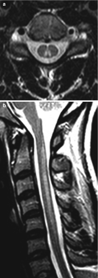
Fig. 11.2
Subacute combined degeneration due to vitamin B12 deficiency. Axial (a) and sagittal (b) MR images show high signal through much of the bilateral cervical spine dorsal columns
Spinal cord infarction: Long-segment T2 hyperintensity and swelling in a spinal artery vascular territory, often involving the central gray matter, are characteristic (Fig. 11.3). Diffusion-weighted imaging is helpful for confirming acute spinal cord infarction by depicting the presence of restricted diffusion in the affected region.
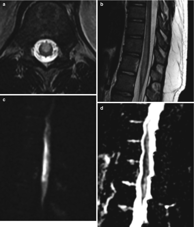
Fig. 11.3
Spinal cord infarction. Axial (a) and sagittal (b) T2-weighted MR images show high signal in the central spinal cord. There is corresponding restricted diffusion shown on the sagittal DWI (c) and ADC map (d)Related posts:

Stay updated, free articles. Join our Telegram channel

Full access? Get Clinical Tree


