Fig. 38.1
Olfactory neuroblastoma. (a) Sagittal T1-weighted precontrast image. (b) Coronal T1-weighted gadolinium-enhanced image. (c, d) Axial fat-saturated T1-weighted gadolinium-enhanced image. There is a large, heterogeneously enhancing mass centered in the floor of the anterior cranial fossa. Nodular components are compressing the frontal lobes and are seen inferiorly within the nasal cavities. The mass extends to the left orbit, with medial displacement of the medial rectus. There is marked edema in the left frontal lobe, and the visualized parts of the lateral ventricles are dilated
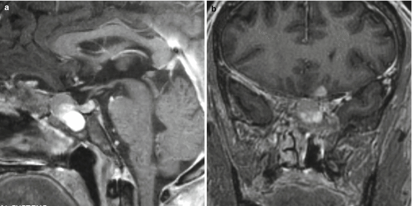
Fig. 38.2
Olfactory neuroblastoma. (a) Sagittal T1-weighted gadolinium-enhanced image. (b) Coronal T1-weighted gadolinium-enhanced image. A heterogeneously enhancing mass is centered in the floor of the anterior cranial fossa, with components eroding the nasal cavities, sphenoid sinuses, and anterior clivus. The pituitary gland is distinctly seen from the mass on the sagittal plane. There is a small cystic component superiorly abutting the left frontal lobe
38.3 Histopathology
Classically, ONBs are characterized by small, round, blue cells slightly larger than mature lymphocytes, with a very high ratio of nucleus to cytoplasm, arranged in a lobular pattern (Fig. 38.3a, b).
ONB is often graded according to Hyams criteria, which range from Grade I to Grade IV [12]. These criteria are based on several features, including architecture, pleomorphism, neurofibrillary matrix, rosettes, mitoses, necrosis, glands, and calcification. Grade I ONB is characterized by lobular cytoarchitecture, little pleomorphism, prominent neurofibrillary matrix, Homer-Wright pseudorosettes, no mitoses or necrosis, and variable calcification. On the other extreme, Grade IV ONB is characterized by loss of lobular cytoarchitecture, marked pleomorphism, necrosis, mitoses, Flexner-Wintersteiner rosettes, and absent calcification [12].
ONBs are typically positive for synaptophysin (Fig. 38.3c), chromogranin, CD56, neuron-specific enolase, neurofilament protein (NFP), and S100 protein [2].
The Ki-67 proliferative index is usually high, ranging between 10 and 50 % [2].
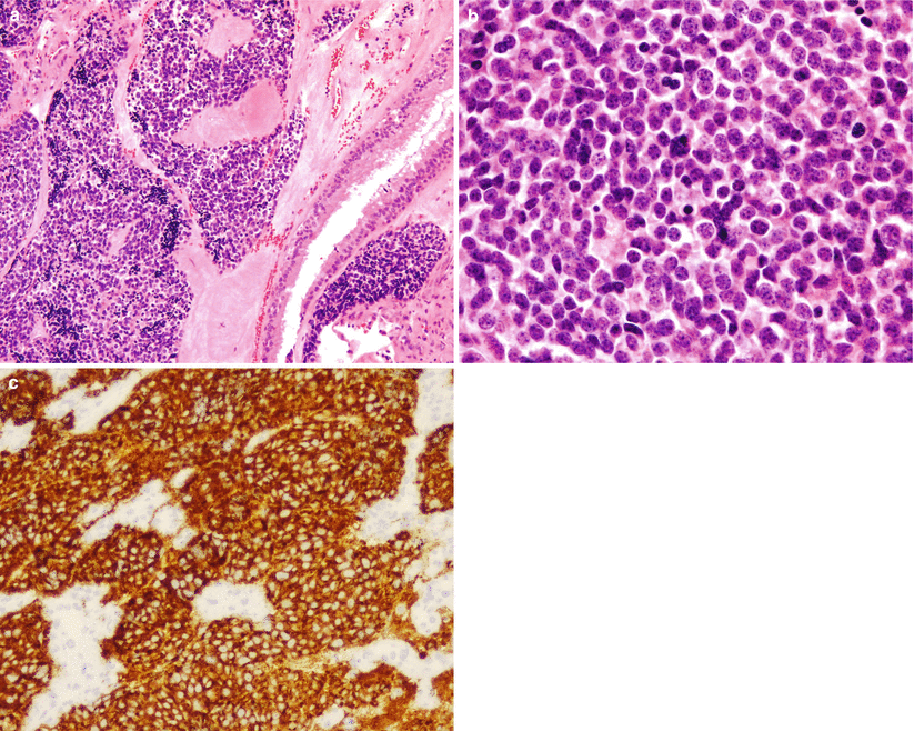
Fig. 38.3
Olfactory neuroblastoma. (a) Olfactory neuroblastomas are located in the sinonasal submucosa. (b) The tumors are composed of a uniform population of small cells with typical “salt and pepper” or dispersed chromatin. Mitotic activity may be high. (c) The tumors are characteristically strongly immunoreactive for synaptophysin
38.4 Clinical and Surgical Management
Depending on the stage of disease at the time of diagnosis and the degree of tumor resection, multimodality treatment consisting of surgery, radiation, and chemotherapy is often implemented.
One large meta-analysis showed no difference in survival between patients who underwent surgery alone and those who underwent surgery plus radiotherapy at 5 years (78 % vs. 75 %) or 10 years (67 % vs. 61 %) [13].
Survival has been associated with Kadish stage and Hyams stage [11, 13]. According to the original Kadish staging, patients with Kadish A tumors had a 5-year survival of 75–91 %, compared with 41–47 % for patients harboring Kadish C tumors [11].
Surgical resection may be performed via standard open craniotomy, craniofacial approaches, or more recently, extended endoscopic endonasal approaches [14].
In a series of 11 patients by Gallia et al. [14], complete endoscopic endonasal surgical resection was possible in all patients, with no patients demonstrating recurrence at 28 months follow-up.
Recent meta-analyses have lent support to equal or more favorable outcomes following endoscopic resection compared with open surgery, although some selection bias exists in the surgical approaches assigned to varying Kadish ONB subtypes [15, 16].
The benefits of adjuvant radiation have been debated; some studies have shown a beneficial effect [17] and others have shown no major benefits [13].Related posts:

Stay updated, free articles. Join our Telegram channel

Full access? Get Clinical Tree


