Optic Pathway Gliomas
Optic pathway gliomas (OPGs) are one of the most challenging neoplasms in modern pediatric neuro-oncology. Recent advances in imaging, surgery, and chemotherapy may lead to a better understanding of the pathophysiology and better clinical results. This chapter reviews these advances and the current treatment paradigms.
36.1 Definition and Classification
The term optic pathway glioma is used to define low-grade glial neoplasms that are epicentered within the visual system: the optic nerve (ON), optic chiasm, optic tracts, and, rarely, the optic radiations. The various presentations of OPG are illustrated in ▶ Fig. 36.1. These lesions tend to have an erratic natural history, and they require careful follow-up and management by a multidisciplinary team. The subset of patients seen by pediatric neurosurgeons may be biased toward the aggressive end of the spectrum, with tumors that have a tendency to progress and require multiple treatment lines.
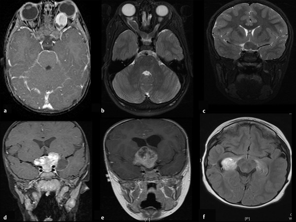
Fig. 36.1 Various presentations of optic pathway glioma. (a) Isolated optic nerve (ON) glioma of the left ON. (b) Bilateral ON glioma involving the chiasm. (c) Chiasmal glioma with no involvement of the hypothalamus. (d) Large chiasmal glioma involving the hypothalamus with a cystic component. (e) Large chiasmatic/hypothalamic glioma that compresses the third ventricle, causing hydrocephalus. (f) Posterior glioma of the optic radiations.
OPGs comprise a wide spectrum of anatomical variations. In addition, age, the coexistence of neurofibromatosis type 1 (NF-1), histology, and molecular markers may be important factors in the clinical behavior of OPGs and in the practical process of individualized decision making regarding their treatment.
Several different methods have been developed for the classification of OPGs. Dodge et al introduced the first of these in 1958.1 The Dodge system classifies OPGs into three groups based on the anatomical location of the tumor (▶ Fig. 36.2). Tumors of the ON are Dodge I, tumors of the chiasm are Dodge II, and posterior tumors, or those that extend into nearby structures, are Dodge III. This method, defined in the era before computed tomography (CT) and magnetic resonance (MR) imaging, is still widely used, mainly for research purposes. However, its clinical relevance is limited.
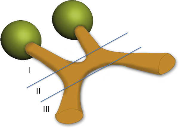
Fig. 36.2 Schematic representation of the 1958 Dodge classification system. I, optic nerve glioma; II, chiasmatic glioma; III, posterior glioma or chiasmatic with involvement of extraoptic structures.
The modified Dodge classification system, developed in 2008,2 is a more detailed anatomical classification that breaks down each component into several highly precise categories. The modified Dodge system also takes into account additional factors, such as the existence of NF or leptomeningeal dissemination.2 This method, although very precise anatomically, is probably too complicated to be routinely implemented in clinical patient care. Another drawback of both Dodge classification methods is the lack of sensitivity to indicate tumor progression.
A third classification system that attempted to address the issue of functional status was suggested by McCullough and Epstein in 1985.3 This classification system consists of two components, tumor/anatomical and functional. The tumor component is divided into four classes (T1, one ON; T2, both ONs; T3, optic chiasm; T4, hypothalamus/thalamus). The functional component is divided into five classes (V0, normal; V1, impaired, one eye; V2, impaired, both eyes or blind, one eye; V3. blind, one eye and impaired, one eye or field defect; V4, blind, both eyes). We recently suggested a new morphological classification that utilizes recent advances in imaging and may aid in clinical management and patient stratification (▶ Table 36.1, ▶ Fig. 36.3). Other factors that should be considered in an attempt to stage an OPG are the presence of NF-1, the age at diagnosis, the symptoms at presentation, and the risk for hydrocephalus.
| Nerve | Chiasm | Posterior | General |
| 1. Mild thickening | 1. Confined to chiasm | 1. Focal involvement | Cyst: yes/no |
| 2. Severe thickening | 2. Chiasm and hypothalamus | 2. Extensive involvement | Hydrocephalus: yes/no |
| Enlarged ONSD: yes/no | 3. Chiasm and third ventricle | Other CNS malignancies: yes/no | |
| Tortuous ON: yes/no | 4. Major suprasellar involvement | Diffuse NF-related changes: yes/no | |
| Pressure on globe: yes/no | Age at presentation? | ||
| Enhancement: yes/no | Enhancement: yes/no | Enhancement: yes/no | Favorable molecular properties? |
| Isolated involvement: yes/no | Isolated involvement: yes/no | Isolated involvement: yes/no | |
| Abbreviations: CNS, central nervous system; ONSD, optic nerve sheath diameter; NF, neurofibromatosis. | |||
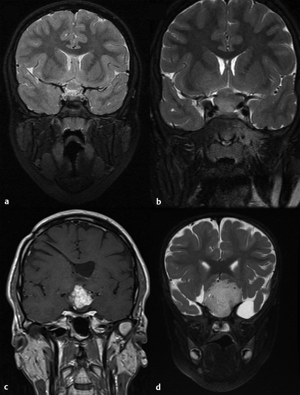
Fig. 36.3 Four different classes of chiasmatic/hypothalamic glioma. (a) Chiasmal thickening only. (b) Chiasm and hypothalamus. (c) Chiasm and third ventricle involvement. (d) Major suprasellar involvement.
Caution is warranted when attempts are made to classify OPGs in patients with NF-1. Despite the fact that this is a relatively common tumor in patients with NF-1, many radiologic abnormalities may complicate the diagnosis. An example of such misleading abnormalities are T2 hyperintensities that sometimes have mass effect, and focal signal changes within the ON that may be either preneoplastic or without any significance.
36.2 Epidemiology
OPGs are the most common primary neoplasms of the neural visual pathways, comprising approximately 1% of all central nervous system (CNS) neoplasms in the general population and approximately 5% of CNS neoplasms in children.4 The annual incidence (as reported in the pre-MR imaging era) is 1 per 100,000.5 The real number is probably higher. Of all patients with this diagnosis, 80% are in the first decade of life, and 90% are in the first two decades. The mean age at diagnosis is 8.8 years in older series and ranges between 2.7 and 5.4 years in more recent series.6–8 An estimated 37% of OPGs tend to progress.9 There is no gender predisposition. In a large series published by Nicolin et al, 58% of 133 patients with OPG were positive for NF-1. The mean age at diagnosis in this series was 5.9 years, 50% of the tumors were hypothalamic/chiasmatic, 60% were diagnosed by imaging only, and 52% required treatment over the course of a follow-up period of 9 years.10 The high proportion of Dodge II or III tumors in this series may be attributable to a referral bias.
36.3 Optic Pathway Glioma and Neurofibromatosis Type 1
OPGs are one of the diagnostic criteria for NF-1, and bilateral OPGs are considered pathognomonic for NF-1. With regard to outcome, the simultaneous occurrence of the two diseases is considered a good prognostic factor.9,11 In a series comparing OPGs associated with NF-1 and sporadic gliomas, the children with NF-1 had a significantly better clinical picture at diagnosis, with less increase in intracranial pressure, less decrease in visual acuity, and fewer abnormalities of the fundus of the eye. Radiologic progression, visual deterioration, and endocrine damage were also less frequent in in children with OPGs associated with NF-1.
OPGs appear in almost 30% of patients with NF-1 and may present with a variety of radiologic and clinical changes.12 In patients with NF-1, the lesion is more likely to be located anteriorly and involve a single nerve than in patients with generic OPG.13 The spectrum of imaging appearances in patients with NF-1 is broad, ranging from fine signal changes on T2-weighted MR images, to an enlarged ON sheath, to gross ON tumors and chiasmatic lesions that protrude into the third ventricle or other neighboring structures. When posterior tumors are being considered in patients with NF-1, it is important to remember that they may easily be confused with NF-related T2 hyperintensities that may be expansile and have a mass effect. A careful radiologic and clinical follow-up is warranted before any therapeutic decisions are made for lesions of the optic radiations. Historically, NF-1 OPGs were considered to be relatively indolent tumors that did not tend to progress. Recently, several publications have described a more active clinical course, with progression of the tumor noted in up to 75% of patients with NF-1, even in children older than 11 years.14 Recently, macrocephaly has also been correlated with OPG in patients with NF-1.15
36.4 Clinical Presentation
Visual complaints such as decreased visual acuity, nystagmus, and proptosis are found at presentation in 46% of patients. Neurologic problems such as headaches, vomiting, and seizures are present in 16% of patients.10 These low percentages are attributed to the young patient age and the high percentage of patients whose tumors are diagnosed during routine screenings, especially patients with NF-1. The age at diagnosis of patients with generic OPGs is older than that of patients with NF-1. This is mainly attributed to the fact that a diagnosis is made only after clinical symptoms appear, not during routine screening, as in patients with NF-1.8
The mode of presentation varies with the anatomical location of the tumor. Posterior tumors are associated with evidence of endocrine dysfunction, such as precocious puberty, and with hydrocephalus. Anterior tumors are associated more with visual abnormalities.16 Visual signs such as palsies of cranial nerves III, IV, and VI, papilledema, and optic atrophy are present in a minority of cases. The type of visual deficit usually correlates with the tumor location; posterior tumors may cause hemianopia, whereas unilateral ON tumors will cause monocular visual impairment. Signs and symptoms of raised intracranial pressure should be taken seriously because acute hydrocephalus has been reported as a presenting symptom. Diencephalic syndrome (cachexia, macrocephaly, nystagmus, and visual deficit) is seen in 21% of infants with chiasmatic/hypothalamic tumors.17
36.5 Diagnostic Studies
36.5.1 Radiology
MR imaging is the gold standard for the diagnosis of OPG. To maximize the diagnostic yield, MR protocols should be planned with the assistance of an experienced neuroradiologist and should include orbit-directed scanning with fat suppression and contrast injection. Tractography may prove beneficial in the future but so far remains experimental.18
Tumors may differ in MR appearance depending on their location, but they are usually isointense on T1 and hyperintense on T2, receive variable enhancement, and have cystic as well as solid nonenhancing components. Tumors of the ON usually do not have cystic changes, appearing as gross thickening of the nerve itself, with or without nerve sheath enlargement. The differential diagnosis for ON enlargement is broad and includes meningioma, neuroma, hemangioblastoma, and lymphoma.19 ON sheath enlargement is found in nonneoplastic conditions such as increased intracranial pressure, optic neuritis, Graves disease, sarcoidosis, toxoplasmosis, central vein occlusion, idiopathic intracranial hypertension, and tuberculosis.20,21 Posterior tumors may appear as minimal chiasmal thickening, or as large masses protruding into the third ventricle with apparent mass effect. Cystic changes are common and are a part of the natural history of the tumor. Neoplastic changes posterior to the lateral geniculate nucleus are rare and are difficult to differentiate from NF-1 T2 hyperintensities. Unfortunately, the initial radiologic appearance does not correlate with the visual prognosis. We have found deteriorating vision in patients with tumors that seem to be stable anatomically, as well as stable vision in patients whose tumors demonstrated structural progression. It has been suggested that dynamic contrast enhancement may correlate with progression, with larger mean permeability values in aggressive tumors.22
Because of the long follow-up required in these patients and the importance of the early detection of changes in the tumor bulk or in its internal components, we recommend the use of volumetric assessments. These measurements, although time-consuming, may improve patient care by enabling more accurate decision making.23,24 ▶ Fig. 36.4 demonstrates a volumetric follow-up of one of our patients; the corresponding MR images are presented in ▶ Fig. 36.5.
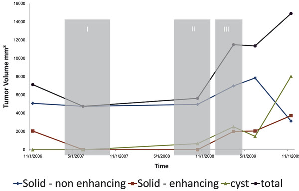
Fig. 36.4 Volumetric follow-up of one of our patients with internal segmentation into three components and integrated treatment periods. Note that despite the fact that the gross total tumor volume enlarges, there is a marked reduction of the solid component following chemotherapy. The main progression is of the cystic component. Total, gross total volume of the tumor; Solid–enhancing, volume of the enhancement-receiving bulk on magnetic resonance (MR) imaging; Solid–nonenhancing, volume of the solid portion of the tumor that does not receive contrast enhancement on MR imaging; Cyst, volume of the cystic component on MR imaging. I, treatment period with vincristine and carboplatin; II, treatment period with vinblastine; III, treatment period with rapamycin and erlotinib (Tarceva; Genentech, South San Francisco, CA).
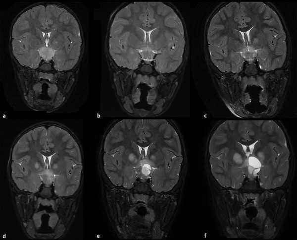
Fig. 36.5 Anatomical history of a chiasmatic/hypothalamic glioma. (a-f) Serial coronal T2 images of a 4-year-old boy with neurofibromatosis type 1 who presented with visual deterioration. Within 4 years, a deterioration to bilateral blindness occurred despite three different chemotherapy treatment lines. (e,f) Note the cystic changes that the tumor undergoes.
36.5.2 Ophthalmology
Neuro-opthalmology examination is crucial for both the diagnosis and follow-up of OPG, as visual decline is a worrisome consequence of the tumor. The neuro-opthalmologic evaluation is often difficult, especially in young patients whose cooperation is limited. A decline in visual acuity is often present at diagnosis and sometimes may be the reason for initial testing, even in the very young (e.g., an infant who starts bumping into objects or sitting closer to the television). In addition, color vision, visual field, eye movements, relative afferent pupillary defect, pupil size, and the fundus should all be evaluated. Any progressive change should be considered seriously as a reason to initiate therapy. In very young children, a normal examination does not rule out visual impairment from an OPG. Recently, the use of optical coherence tomography (OCT) was shown to detect loss of the retinal nerve fiber layer in children with OPG (▶ Fig. 36.6). This may prove to be an auxiliary tool in diagnosing visual damage in young children, as well as in providing evidence regarding visual reserve and the need for treatment.25,26
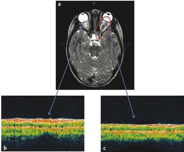
Fig. 36.6 Patient with neurofibromatosis type 1 and optic pathway glioma (a) with a tumor on the left optic nerve (red circle). Optical coherence tomography of the right, normal eye (b) and of the left, tumor-affected eye (c) (blue arrows). This patient had a visual acuity of 20/20 in the unaffected (right) eye and of 20/400 in the tumor-affected eye. Visual acuity correlated well with retinal nerve fiber layer thickness of 105 µm on the right (normal) versus 48 µm on the left (severely thinned).
(Images courtesy of Dr. Robert Avery, Children’s National Hospital, Washington. DC.)
36.5.3 Endocrine Assessment
An endocrine assessment should be done in any child with chiasmatic/hypothalamic glioma. Endocrine abnormalities such as central precocious puberty and growth hormone deficiency are detected in approximately 20% of patients with an OPG. It should be noted that growth hormone deficiency is present in approximately 2.5% of patients with NF-1, even those without OPG.27
36.6 Pathology
OPGs are typically low-grade glial neoplasms. Pilocytic astrocytoma accounts for the vast majority of these tumors. The rest usually consist of fibrillary and pilomyxoid astrocytomas, oligodendrogliomas, and gangliogliomas. A more aggressive behavior pattern may be predicted with labeling indices such as high Ki-674,28 as well as pilomyxoid histology.29,30 Malignant transformation is rarely seen and is most frequently associated with irradiation.31
36.7 Natural History and Prognosis
OPGs have an erratic natural history. Some of these tumors progress, others are steady for a lifetime, and others spontaneously regress.32,33 This has led to the theory that OPGs are not a single entity, but rather a group composed of two or even three subtypes that are often hard to distinguish from one another. These subtypes vary widely in their clinical and radiologic outcomes. Although some OPGs undergo spontaneous regression, a significant group of large chiasmatic/hypothalamic tumors tend to progress and even metastasize. ▶ Fig. 36.4 demonstrates a progressive OPG of the chiasmatic/hypothalamic type over 4 years of follow-up. Interestingly, of the 5% of low-grade gliomas that undergo leptomeningeal spread, OPGs account for approximately 50%.34 Before this behavior pattern was recognized, these tumors were sometimes considered benign, even hamartomatous in nature,35 requiring only careful follow-up; however, they are now perceived as progression-prone, persistent tumors that pose a major therapeutic dilemma. As many as 35% require treatment at presentation.10
Patients who do not require treatment at initial presentation have varying chances for progression. In those with NF-1, the likelihood of progression is 15%, whereas in patients with sporadic OPGs, the probability of progression is 75%.
Molecular analysis of tumor tissue has shown promising early results in predicting the course of the disease. In particular, the BRAF-KIAA 1549 fusion protein seems to indicate tumors with a tendency to arrested growth and even to spontaneous senescence. In a recent study, the 5-year progression-free survival (PFS) rate was 61% ± 8% for B-K fusion protein–positive patients and 18% for B-K fusion protein–negative patients. In this study, 61% of sporadic OPGs were positive for fusion protein, and no OPG related to NF-1 was positive for this mutation.36 Although not currently utilized in routine clinical practice, we believe that this analysis should be considered for any patient without NF-1 for whom therapeutic decisions are being made based on radiologic progression only.
36.8 Treatment
The follow-up and management of OPG require an experienced multidisciplinary team that provides individualized patient care. Only then can adequate therapeutic decisions be made. The goal of treatment is to prevent visual decline and to achieve long-term tumor control. In the presence of a severe mass effect or hydrocephalus, immediate, life-saving neurosurgical procedures may be indicated. In most cases, however, OPGs are not life-threatening tumors. The risk-to-benefit ratio of treatment must therefore be considered carefully for each patient.
The exact timing of treatment initiation is one of the major open questions in OPG management. In most cases, we recommend delaying treatment for as long as possible unless a clear radiologic progression or visual decline is noted. Treating an asymptomatic or minimally symptomatic stable patient seems to offer no advantage over observation alone.6,28,37 Thus, current guidelines suggest intervention only when there is a documented decline in vision or radiologic progression.9 Treatment initiation dilemmas may be very relevant in a child, especially an infant, who presents for the first time with compromised vision. At this moment, progression cannot be defined; however, compromised reserve may be a relevant reason to start treatment earlier rather than later.
We recommend imaging and neuro-ophthalmologic examinations (including visual field and OCT if available) every 6 months. Patients with compromised reserve for whom a decision has been made not to treat should be clinically examined more frequently. If the tumor is chiasmatic/hypothalamic, a detailed endocrinologic evaluation should be performed. If the patient has been stable for a period of a year and has no adverse prognostic factors, follow-up visits may be scheduled on a yearly basis.
The choice between treatment options such as surgery, chemotherapy, and irradiation is not easy and depends on the team’s experience and biases. The following sections outline general rules for these treatment modalities. ▶ Fig. 36.7 illustrates a suggested management algorithm for OPG.
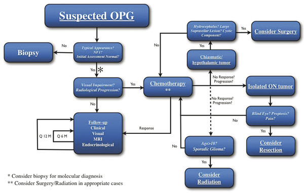
Fig. 36.7 Management algorithm for patients with optic pathway glioma. MRI, magnetic resonance imaging; ON, optic nerve; OPG, optic pathway glioma. See text for specifics.


