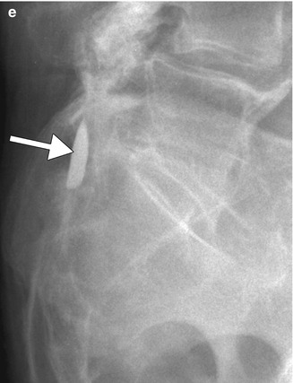
Fig. 14.1
Intraspinal Pantopaque. Axial and sagittal T1-weighted and T2-weighted MR images (a–d) show high T1 and intermediate T2 signal with associated chemical shift artifact within the spinal canal (arrows). Lateral radiograph of the lumbar spine (e) shows a high attenuation material in the spinal canal at the level of L5-S1 (arrows)

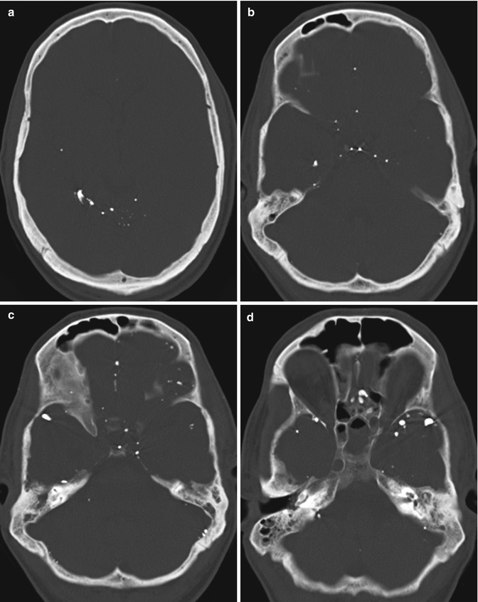
Fig. 14.2
Intracranial Pantopaque. Axial CT images (a–d) show numerous scattered punctate hyperattenuating foci of residual Pantopaque in the subarachnoid spaces
14.4 Differential Diagnosis
In the spine, the differential diagnosis for subarachnoid T1 hyperintense lesions on MRI includes fibrolipomas, lipomas, epidural lipomatosis, dermoid, hemorrhage, and melanoma. Without an appropriate clinical history, CT can help differentiate among some of these possibilities; fat is typically very hypoattenuating on CT, while subacute hemorrhage and melanoma have intermediate attenuation. The differential diagnosis for leptomeningeal dissemination of Pantopaque into the intracranial compartment with hyperattenuation on CT mainly includes meningoangiomatosis and subarachnoid hemorrhage. On MRI, ruptured dermoid, leptomeningeal melanoma and/or hemorrhagic metastases, and subacute subarachnoid hemorrhage are potential differential diagnostic considerations.
Get Clinical Tree app for offline access

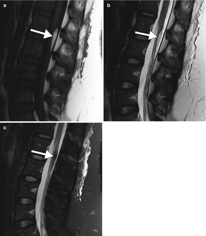
Fibrolipoma: Filum terminale fibrolipomas typically appear as elongated masses with fat signal characteristics (T1 and T2 hyperintense with loss of signal on fat-suppressed sequences) on MRI (Fig. 14.3).

Fig. 14.3
Fibrolipoma of the filum terminale. Sagittal T1-weighted (a), T2-weighted (b), and fat-suppressed T2-weighted (c) MR images show a fusiform mass associated with the filum terminale with signal characteristics of fat (arrows)
Stay updated, free articles. Join our Telegram channel

Full access? Get Clinical Tree


