• The end plate is clearly visualized. The spinous process is centered between both pedicles. The Jamshidi needle is started at the 2 o’clock position.

• The Jamshidi trocar is advanced 15 mm until it is centered in the pedicle.
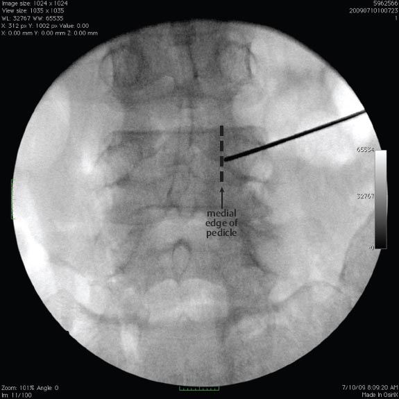
• A guide wire is then advanced an additional 10 mm until it abuts the medial wall of the pedicle on the AP image.
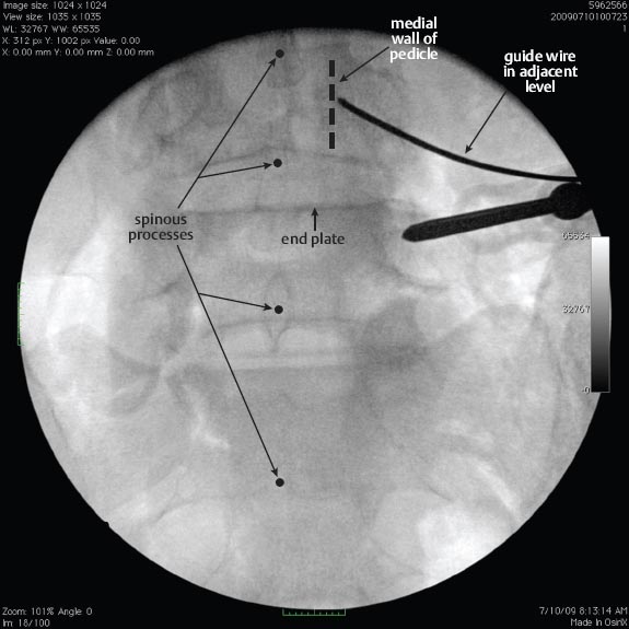
• The steps are repeated for the adjacent level. End plate visualization and centering the spinous process are essential steps for ensuring accurate percutaneous screw placement.

Related posts:

Stay updated, free articles. Join our Telegram channel

Full access? Get Clinical Tree


