| 173 | Percutaneous Retrograde Femoral Artery Puncture and Other Access Techniques |
♦ Preoperative
Special Equipment
- Sterile drapes
- Local anesthetic
- No. 11 scalpel
- Hemostat
- Puncture needle (18-gauge Pott’s or modified Pott’s needle, or a 21-gauge micropuncture needle or kit)
- 0.035-in guide wire
- Dilators (for sheaths larger than 6.0 French [F])
- Sheath (usually 5.0 to 6.0 F)
- Nylon or silk suture
- Scissors
- Sterile 4 × 4 gauze
- Various catheters as needle to negotiate the patient’s anatomy, usually starting with a 5F hockey stick or vertebral catheter
♦ Intraoperative
Positioning
- The patient is placed in supine position on the angiography table
- Intravenous (IV) antibiotics, if needed, are given
- A Foley catheter is placed (for interventional procedures)
- The proper shielding is placed on the patient
- Both inguinal areas are shaved and prepped with iodine solution
- A sterile drape (with adhesive cutouts for the groin) is placed
- The head is placed in neutral position and gently taped in place
Technique
- The femoral artery is palpated with the left hand
- The inguinal ligament is identified (extending between the pubic tubercle and anterior iliac spine)
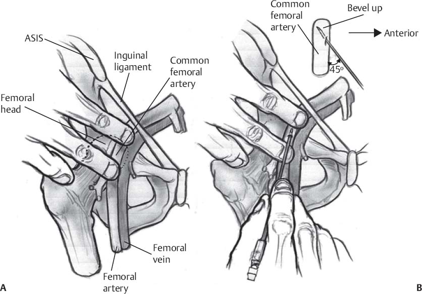
- The path of the common femoral artery (CFA) is gently traced inferior to the inguinal ligament, and is trapped by gently compressing the artery onto the femoral head with the index and middle fingers separated by several centimeters (Fig. 173.1A)
- The region between the index and middle fingers (~3 cm inferior to the inguinal ligament) is infiltrated with local anesthetic.
- A small (3 to 4 mm) skin incision is made.
- The underlying soft tissue is dissected with a small hemostat to create the dermatotomy tract.
- The tip of the puncture needle is directed cranially at a 45-degree angle parallel to the course of the CFA and placed in the dermatotomy tract (Fig.173.1B).
- If the needle is directed correctly, the pulsatility of the femoral artery will be transmitted along the needle with the tip of the needle resting on the vessel wall.
- Either a single- or double-wall arterial puncture is performed.
- Single wall technique
- The Pott’s needle is firmly and smoothly advanced through the anterior wall of the artery
- The inner stylet is removed to assess the degree of back-bleeding
- If back-bleeding is not vigorous, the inner stylet is gently replaced and the Pott’s needle is advanced a little deeper; the stylet is again removed
- Once brisk, pulsatile back-bleeding occurs, the soft, floppy portion of the guide wire is advanced through the cannula
- If resistance is felt, the wire is withdrawn and the tip of the cannula redirected slightly (the wire might have been against the vessel wall)
- Once the wire advances easily, the cannula is gently withdrawn over the wire with the operator’s right hand until the wire near the skin can be secured with the left hand; the cannula is then completely removed from the wire
- The Pott’s needle is firmly and smoothly advanced through the anterior wall of the artery
- Double wall technique
- The Pott’s needle is firmly and smoothly advanced through the artery down to bone, thus traversing both arterial walls
- The stylet is removed, and the Pott’s cannula is slowly retracted until brisk pulsatile back-bleeding occurs
- The soft, floppy portion of the guide wire is advanced through the cannula
- If resistance is felt, the wire is withdrawn and the tip of the cannula redirected slightly (the wire might have been against the vessel wall)
< div class='tao-gold-member'> Only gold members can continue reading. Log In or Register to continue
Stay updated, free articles. Join our Telegram channel
- The Pott’s needle is firmly and smoothly advanced through the artery down to bone, thus traversing both arterial walls

Full access? Get Clinical Tree


