Photic Stimulation Responses
▲ Photic Driving Response
Other Names
Occipital driving response
Description
Visual stimulation with a flashing strobe according to certain standard parameters commonly produces a run of sharply contoured, positive, monophasic transients that are called a photic driving response. Usual stimulation parameters include strobe frequencies between 1 and 30 Hz for periods lasting 5 to 10 seconds (Kasteleijn-Nolst Trenite et al., 2012). Each transient follows a flash by 80 to 150 milliseconds and the transients usually recur at the same frequency as the stimulation. However, the run of transients can instead be a harmonic or a subharmonic of the stimulation frequency. Photic driving responses are best elicited with red light or with the eyes closed, which produces a red filtering of the strobe light as it passes through the eyelids, but periods with eyes opened also are performed (Takahashi, 1999). The stimulation frequency that is most likely to produce a response varies with age and usually is close to the frequency of the individual’s alpha rhythm, but it is not always exactly the same frequency (Kiloh et al., 1981). The photic driving response’s amplitude is maximal with stimulation frequencies close to the alpha rhythm’s frequency and decreases with greater differences between the stimulation frequency and alpha rhythm frequency.
The photic driving response may be seen at ages as young as 3 months, and usually first appears around the age at which the alpha rhythm first develops. However, the alpha rhythm in infants and young children is not within the alpha frequency range, and it is commonly called a posterior dominant rhythm for this reason. The posterior dominant rhythm is in the theta range when it first occurs, and the frequencies that elicit the photic driving response are similarly slow. When the posterior dominant rhythm reaches the alpha frequency range at around the age of 3 years, the photic driving response also reaches the alpha frequency range (Niedermeyer, 1999c). Overall, photic driving responses may occur at frequencies as broad as from 5 to 30 Hz, which include responses in early life and responses that are subharmonics and harmonics. Moreover, stimulation frequencies less than 3 Hz rarely produce a driving response (Fisch, 1999).
The field of the photic driving response is mainly bilateral occipital but may extend to include the posterior temporal regions. Extension to more anterior regions is uncommon but occurs. The driving response’s amplitude is low until ages of about 6 years and is similar to that of the alpha rhythm from young to middle adulthood. In later adulthood, the amplitude decreases. Also similar to the alpha rhythm, photic driving responses may be asymmetric with greater amplitude on the right (Blume et al., 2002).
Distinguishing Features
• Compared to Lambda Waves
Morphologically, photic driving responses at low frequencies are similar to lambda waves and appear especially similar if the driving response occurs inconsistently, such that the transients do not have a fixed interval. Identifying transients as a photic driving response depends on notation on the EEG record that indicates the photic stimulation frequency and the moments that the strobe flashes. Varying intervals between the waves at different portions of the EEG, as occurs with the photic driving response as the stimulation frequency is changed, also aids in differentiation.
• Compared to Photomyogenic Response
Although the photomyogenic response usually has an anterior field, occasionally it includes head movements that produce posterior artifact from contact between the head and the surface below it. Since the posterior artifact is due to movement and not individual motor unit potentials, they may be blunt and resemble the photic driving response waveform. Furthermore, the movements are time-locked with stimulation like the photic driving response. Differentiation depends on the consistency of the waveform and field. Neither the waveform nor the field
of the driving response varies during stimulation, even across multiple stimulation frequencies, whereas, the photomyogenic response movement artifact’s waveform and location depend on head movements, which are less consistent.
of the driving response varies during stimulation, even across multiple stimulation frequencies, whereas, the photomyogenic response movement artifact’s waveform and location depend on head movements, which are less consistent.
• Compared to Interictal Epileptiform Discharges
When the photic driving response is a harmonic of the stimulation frequency, the elicited transients have the duration of spikes. For example, a 12-Hz stimulation can produce a 24-Hz driving response, which would have individual waves within the response that are 42 milliseconds and appear similar to monophasic epileptic spikes or paroxysmal fast activity. The absence of after-going slow waves is a straightforward differentiating feature from diphasic and triphasic epileptiform discharges. However, either identifying a rhythm to the waves or harmonic relationship with the stimulation is a more reliable means to differentiate these patterns.
• Compared to Photoparoxysmal Response
The photoparoxysmal response usually has a frequency that is less than the stimulation frequency and does not vary with changes to the stimulation frequency, so it does not have a subharmonic relationship. Furthermore, continuation of the response beyond the stimulation period is common and highly differentiating because the driving response ends with cessation of stimulation. The pattern’s field also often differs; the driving response is posterior and the photoparoxysmal response usually is anterior, central, or generalized; however, posterior photoparoxysmal responses can occur.
Co-Occurring Patterns
When photic stimulation is performed during wakefulness with eyes closed, an alpha rhythm is present within the region of the driving response. Frontal artifact that is synchronized with the stimulation also may be present. This artifact may be produced by both an electromyographic response and a electroretinographic effect.
Clinical Significance
A bilateral photic driving response at a frequency close to the patient’s alpha rhythm is a normal phenomenon, but its presence is not necessary for an EEG to be considered normal. The absence of a photic driving response is a common normal variant that is especially common in the elderly (Fisch, 1999). Medications may affect whether a photic driving response is present with adrenergic blocking drugs augmenting the response and adrenergic stimulants diminishing it (Kerxhalli et al., 1975; Vogel et al., 1974).
A photic driving response at a stimulation frequency less than 3 Hz may be abnormal with significance similar to abnormal slowing and it is associated with degenerative encephalopathies, including the Bielschowsky–Jansky form of neuronal ceroid lipofuscinosis, progressive myoclonus epilepsies, and prion disease (Mendez and Brenner, 2006; Naidu and Niedermeyer, 1999). A driving response at a frequency greater than 20 Hz has been associated specifically with migraine (Niedermeyer, 1999a). Asymmetry to the photic driving response is not necessarily abnormal and should be compared to the symmetry of other EEG features and especially the alpha rhythm. If the driving response’s asymmetry is consistent with the normal asymmetries in other features, then it most likely also is normal. In the absence of another asymmetry, asymmetric photic stimulation responses may be a sign of abnormality. However, asymmetric responses also may be due to gaze deviation away from the strobe or atypical calcarine cortex anatomy with a subsequent abnormal dipole orientation. Moreover, abnormal asymmetry does not indicate which side is abnormal. Decreased amplitude may result from destructive lesions, and increased amplitude may result from irritative ones. An abnormal unilateral loss of a photic driving response is more easily interpreted and indicates pathology ipsilateral to the side without the response.
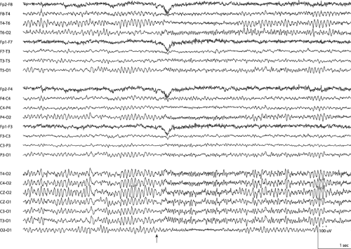
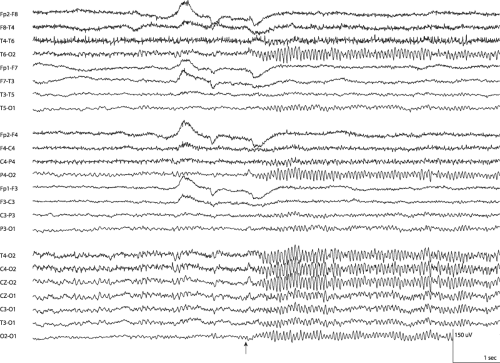 Figure 23-1 Photic Driving Response • Photic stimulation at 14 Hz begins where marked and produces a 14-Hz bioccipital driving rhythm. The amplitude has a minor asymmetry with higher voltage on the right. The asymmetry is present in both short interelectrode and long interelectrode channels, so is not due to a broader field across the left occiput. Blink artifact precedes the stimulation. (LFF 1 Hz, HFF 70 Hz) |
Related posts:Stay updated, free articles. Join our Telegram channel
Full access? Get Clinical Tree
 Get Clinical Tree app for offline access

|


