Physiology of the Intervertebral Disc
Jing Yu
Thijs Grünhagen
Cornelia Neidlinger-Wilke
Sally Roberts
Jill Urban
The intervertebral disc has only a small number of cells embedded throughout a dense extracellular matrix (ECM), which the cells synthesize and maintain. As the mechanical functioning of the disc is governed by the composition and organization of this matrix, the continuing health and activity of these cells is vital for sustaining disc health and its biomechanical role. Here we review recent information on disc cell function and viability, particularly in relation to the response of the cells to extracellular signals such as mechanical stress and nutrient supply. New cell-based therapies proposed for treating disc degeneration are also discussed briefly.
Disc Composition
The ability of intervertebral discs to fulfill their main mechanical tasks (viz. to support compressive loads and act as joints of the spinal column, allowing flexion, torsion, and extension) depends primarily on the organization and composition of the major macromolecules that make up this tissue, these being collagen and the large aggregating proteoglycan, aggrecan (51). The fibrillar collagens form an elaborate network that provides the structural framework of the disc, and aggrecan has a high swelling pressure, imbibes water, and inflates the collagenous network; together these components form a load-bearing structure, allowing the disc to carry compressive load and to deform reversibly (36).
Other macromolecules, although present in only low concentrations, also appear to have essential roles in regulating matrix organization. For instance, elastin, although present in only minor amounts, appears to have an important mechanical function in restoring disc organization after load-induced deformation. The role of other minor components is also important in tissue function and organization; small proteoglycans such as fibromodulin and decorin control the diameter of the fibrillar collagens (19), whereas the minor collagen, collagen IX, appears to play a vital part in linking network structures together (15). It is thus not surprising that polymorphisms in these molecules are associated with disc degeneration (1,15). This information shows that the biomechanical role of the disc is governed by a complex macromolecular network, comprised not only of the major fibrillar collagens and of aggrecan but also of a large number of other proteins and glycoproteins.
Disc Composition Varies With Region
The proportion, composition, and organization of these molecules varies considerably from one region of the disc to the other. The outer region of the disc, the annulus fibrosus, is highly organized (Fig. 3.1). It has a high concentration of fibrillar collagens, organized into concentric lamellae encircling the disc. These lamellae consist of bundles of collagen fibers running obliquely from one vertebral body to the next, firmly anchoring the disc to the bone. The angle of the collagen bundles alternates between successive lamella, thus forming a cross-woven and reinforced structure. Elastic fibers (Fig. 3.1) are concentrated between the annulus lamellae and may play a mechanical role in maintaining annulus organization. The annulus encloses the softer, more hydrated nucleus pulposus, which by contrast has only a loose random collagen network but has a high concentration of aggrecan compared to the annulus or to most other cartilages (51). The disc is separated from the vertebral body by a thin (1 mm) layer of hyaline cartilage, the cartilaginous endplate, which is less hydrated than the adjacent disc regions.
Changes With Age and Pathology
There are large visible changes in appearance of the disc with age and degeneration. With increasing age, the nucleus becomes less hydrated and more collagenous; it discolors, changing from white and translucent in youth to becoming yellow-brown
and opaque, and the boundary between nucleus and annulus becomes blurred (53). The annular rings thicken and appear to lose organization (Fig. 3.1). Eventually cracks and fissure appear in the endplate, nucleus, and annulus and the disc thins and distorts. These age- or degeneration-linked changes parallel (or follow) changes in disc composition. One of the most evident age- and/or degeneration-related changes is loss of aggrecan (Fig. 3.2). Because aggrecan, on account of its high swelling pressure, is responsible for maintaining disc hydration, loss of hydration follows from loss of aggrecan. Changes in the minor macromolecules have been less well studied, but recent work has shown that small proteoglycan concentrations in the nucleus also fall with increase in degeneration (13).
and opaque, and the boundary between nucleus and annulus becomes blurred (53). The annular rings thicken and appear to lose organization (Fig. 3.1). Eventually cracks and fissure appear in the endplate, nucleus, and annulus and the disc thins and distorts. These age- or degeneration-linked changes parallel (or follow) changes in disc composition. One of the most evident age- and/or degeneration-related changes is loss of aggrecan (Fig. 3.2). Because aggrecan, on account of its high swelling pressure, is responsible for maintaining disc hydration, loss of hydration follows from loss of aggrecan. Changes in the minor macromolecules have been less well studied, but recent work has shown that small proteoglycan concentrations in the nucleus also fall with increase in degeneration (13).
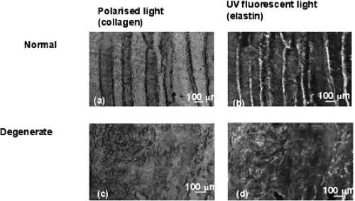 FIGURE 3.1 The organized lamellar structure of normal discs is lost in degenerated discs. The figure shows sections from normal adult annulus (A, B) and degenerate (grade 2–3) adult annulus from the same region (C, D). The sections are shown under polarized light to visualize collagen organization (A, C) and under fluorescent light to visualize elastin (B, D). |
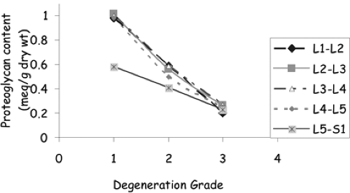 FIGURE 3.2 Loss of proteoglycan with degeneration grade. Changes in proteoglycan content, measured in relation to concentration of negatively charged groups/dry weight, variation with spinal level, and disc degeneration grade (adapted from reference 51). |
Matrix Degrading Enzymes
As well as the macromolecular components discussed previously, the disc also contains several different classes of proteases, that is enzymes that can degrade matrix proteins (30,34,41,42,53). Inhibitors, which block activity of these enzymes, are also present in the disc. In their active form, that is, when the inhibitors are nonfunctional, these proteases can break down and disaggregate most of the macromolecular components of the disc. With age and pathology the level of active proteases present in the disc increases, supporting their suggested role in disc matrix turnover and degradation (11,41).
Disc Cells
The matrix is maintained and turned over by a small population of resident cells whose main function is to synthesize and maintain an appropriate macromolecular composition. The disc cells produce matrix macromolecules throughout life. They also produce
proteases and their inhibitors. The disc remains healthy while the rate of macromolecular synthesis and breakdown is in balance; however, if the rate of breakdown increases over synthesis, ultimately the disc matrix begins to disintegrate. Disc degeneration thus results from failure of the disc’s cells to produce, maintain, and repair the matrix.
proteases and their inhibitors. The disc remains healthy while the rate of macromolecular synthesis and breakdown is in balance; however, if the rate of breakdown increases over synthesis, ultimately the disc matrix begins to disintegrate. Disc degeneration thus results from failure of the disc’s cells to produce, maintain, and repair the matrix.
Cell Types in the Disc
Relatively little is known of the cells of the disc. Cell density is low, being around 0.25% to 0.5% of tissue volume and varies inversely with disc thickness in a manner that suggests it is under nutritional control (47). The cells of each region of the disc appear phenotypically distinct, possibly reflecting different embryologic origins (Fig. 3.3). The nucleus is derived from the notochord. Notochordal cells remain in the center of the disc for only a short time span in humans, but in other mammals (e.g., rodents) they are retained into old age (8). The nucleus pulposus of the adult disc is populated by what appear to be round/oval cells, resembling chondrocytes of cartilage (Fig. 3.3), often lying within a microfibrillar capsule. With aging or degeneration, nucleus cells can proliferate to form clusters (28). The annulus derives from the mesenchyme. Its highly organized ECM is dependent on the initial orientation of the cells directed by their stress fiber and cytoskeletal organization (18). The cells of the annulus are often
elongated, especially in the outer region, and orientated along the lamellae (7); they can be described as fibroblastlike. The annulus and nucleus cells have extended cell processes, forming a network of cell-cell and cell-matrix contacts whose role is unknown but is thought to be related to mechanotransduction (7,14,27,37).
elongated, especially in the outer region, and orientated along the lamellae (7); they can be described as fibroblastlike. The annulus and nucleus cells have extended cell processes, forming a network of cell-cell and cell-matrix contacts whose role is unknown but is thought to be related to mechanotransduction (7,14,27,37).
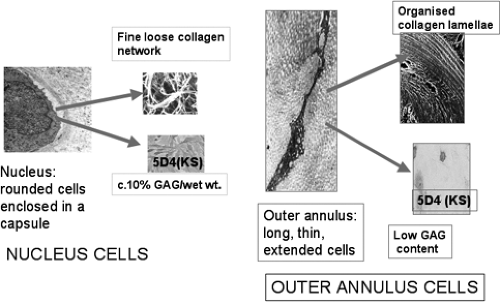 FIGURE 3.3 Cells of the intervertebral disc. Figure showing differences in cell phenotype and matrix produced (adapted from references 14 and 22). Rounded cells from the nucleus produce a high concentration of GAGs (immunostained brown with antibody to keratan sulphate, 5D4) and a loose collagen network. Extended fibroblastlike cells from the outer annulus produce low concentrations of GAG and highly organized collagen lamellae. |
Related posts:
 Radiologic Assessment of All Unfused Lumbar Segments After Instrumented Posterior Spinal Fusion
Natural Evolution and Resolution of Chronic Low Back Pain
Lumbar Surgical Approaches for Nonfusion Techniques
Nucleus Pulposus Replacement: Basic Science and Indications for Clinical Use
Influence of Frontal and Sagittal Position of Total Disc Arthroplasty on Clinical Outcomes at 3 Years Follow-Up
Posterior Nonfusion Stabilization of the Degenerated Lumbar Spine with Cosmic
Radiologic Assessment of All Unfused Lumbar Segments After Instrumented Posterior Spinal Fusion
Natural Evolution and Resolution of Chronic Low Back Pain
Lumbar Surgical Approaches for Nonfusion Techniques
Nucleus Pulposus Replacement: Basic Science and Indications for Clinical Use
Influence of Frontal and Sagittal Position of Total Disc Arthroplasty on Clinical Outcomes at 3 Years Follow-Up
Posterior Nonfusion Stabilization of the Degenerated Lumbar Spine with Cosmic
Stay updated, free articles. Join our Telegram channel

Full access? Get Clinical Tree


