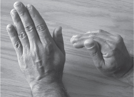
33 Posterior Interosseous Nerve Injury An otherwise healthy 58-year-old man presented elsewhere with right lateral elbow pain radiating into the ulnar aspect of the hand. He underwent a decompressive procedure of the right lateral epicondyle and posterior interosseous nerve (PIN). There was slight but incomplete resolution of the pain and he underwent a second operation 1 year later through a posterolateral incision. Upon awakening in the recovery room, he was unable to extend his fingers. There was no change in his pain symptoms. Over the following 2 months, he did not experience any improvement in his finger drop. Upon examination in our clinic there was normal function of triceps and brachioradialis. Wrist extension occurred with radial deviation, revealing 5/5 power in the extensor carpi radialis but 0/5 power in the extensor carpi ulnaris. There was 0/5 power in the finger extensors, abductor pollicis longus, and extensor pollicis longus. Sensory examination over the dorsal aspect of the forearm and wrist was normal. There was no Tinel sign. Electromyographic examination showed fibrillation potentials and positive waves in the extensor carpi ulnaris and extensor communis, consistent with denervation. Nerve conduction studies of the radial and ulnar nerves were normal. One month later, with no clinical improvement, he underwent surgical exploration of the right radial and posterior interosseous nerves. An anterior incision was made along the biceps brachialis muscle medially and the brachioradialis muscle laterally, and the radial nerve was examined along its length. The radial nerve was normal to the level of the PIN, as was the superficial sensory branch of the radial nerve. At the level of the supinator muscle, a neuroma in continuity was seen that extended around the supinator muscle and to the level of the branches of the PIN as they entered their respective extensor compartment muscles. The distal part was exposed through an incision made posteriorly extending along the initial incision line. Intraoperative electrical studies demonstrated a lack of electrical continuity of the nerve through the neuroma. A 5 cm length of PIN was removed containing the neuroma in continuity, and was repaired with a superficial sensory radial nerve graft fashioned into two cables. Over the next year he experienced gradual improvement in his ability to extend his wrist and fingers, but he had continuing pain over the extensor muscle mass, which responded partially to nonsteroidal anti-inflammatory medications and tricyclic antidepressants. Two years post-operatively he had 4/5 power in the extensor carpi ulnaris and finger extensors and nearly full function of the hand, but he was still affected by ongoing pain over the dorsal aspect of the forearm. Posterior interosseous nerve injury The radial nerve in the arm courses around the humerus and pierces the lateral intermuscular septum. In the lateral arm it lies between the brachialis and brachioradialis muscles and enters the antecubital fossa under the cover of the brachioradialis and extensor carpi radialis longus. The main radial nerve gives motor supply to the brachioradialis and extensor carpi radialis longus 2 to 3 cm proximal to the elbow. The nerve to the extensor carpi radialis brevis is also a separate nerve that originates from the main radial nerve near its bifurcation. The radial nerve divides into the superficial sensory radial nerve (which descends in the forearm under the edge of the brachio-radialis and lateral to the radial artery) and PIN 1 to 2 cm distal to the lateral epicondyle, but this may be variable. The fascicles destined for the PIN are located more posteriorly in the radial nerve at this level. The PIN supplies all of the extensor muscles of the back of the forearm except the extensor carpi radialis longus. The PIN spirals around the radius between the two heads of the supinator muscle and supplies the supinator and extensor carpi ulnaris before it passes between the heads of the supinator muscle. The volar supinator forms an arch around the PIN that is fibrous in 30% of people and here is called the arcade of Frohse. Upon exiting the distal border of the supinator, the PIN divides into short branches that supply the medial extensor musculature (extensor carpi ulnaris, extensor digitorum communis, extensor digiti minimi) and two long branches that supply the extensor pollicis longus, extensor pollicis brevis, abductor pollicis longus, and extensor indices. There is some sensory supply by the PIN to the ligaments and joints about the carpal bones, and to the periosteum of the radius, but no cutaneous innervation is derived from it. Our patient typifies the clinical syndrome of PIN injury. There is weakness of the muscles supplied by the PIN, with sparing of those supplied more proximally by the radial nerve. Thus the weakness involves the finger extensors and the extensor carpi ulnaris and spares the triceps, brachioradialis, supinator, and extensor carpi radialis. The patient is able to extend the interphalangeal joints with the intrinsic muscles of the hand. Essentially, the patient with a pure PIN palsy has a finger drop, with wrist extension (in a radial direction) largely spared (Fig. 33–1). There is no sensory deficit, and pain is not a major feature of this syndrome, although patients may have a dull aching sensation over the forearm, especially in cases of entrapment. In cases of entrapment, the motor loss in the distribution of the PIN can be more variable and is often less complete than with injury. In terms of etiology, PIN palsy may be either traumatic or nontraumatic. In Young et al’s study of 40 PIN palsies treated surgically, the injury was iatrogenic in 15, traumatic in 16, and nontraumatic in nine. Of the iatrogenic injuries, nine involved operations of the proximal radius. In another study by Cravens and Kline, of 32 PIN palsies, 15 were traumatic (lacerations in six, fractures in three, operative repair of radial fractures in three, and three contusions) and 17 nontraumatic (14 entrapments and three tumors). Most iatrogenic injuries result from operations involving the proximal radius but also include operations for release of PIN entrapment or for tumor resection in that area. The patient presented with pain initially. The initial symptoms may have represented radial tunnel syndrome or lateral epicondylitis, both of which involve aching pain over the lateral elbow, without motor deficits. They are described elsewhere in this volume (Chapter 32 on radial tunnel syndrome). Our patient, having symptoms immediately post-operatively, clearly had iatrogenic injury to the PIN. The major differential diagnosis in such a case involves which nerve is injured and at what anatomical level. This relates to the anatomy of the radial nerve, as already described. It is recommended that the decision whether to operate be made by 3 months postinjury. The exception is that patients with sharp injuries such as lacerations are candidates for early repair. In general, with injuries in continuity, one should wait to ensure that there is no early recovery (as would occur with a neurapraxic injury), as demonstrated by lack of clinical improvement and no electrophysiological evidence of recovery. The clinical picture is often more reliable than electrophysiological studies. In Young et al’s series, of the 51 patients with PIN palsy, 11 resolved without surgical treatment. The PIN may be approached from an anterior incision, a posterior incision, or a combination of both. In one study of 40 operatively treated PIN lesions, 16 required an anterior approach, 23 a combined approach, and one a posterior approach. The operative treatment options are external neurolysis and release of entrapment; internal neurolysis; resection of neuroma in continuity with primary repair or graft; and tendon transfers.
 Case Presentation
 Diagnosis
 Anatomy
 Characteristic Clinical Presentation
 Differential Diagnosis
 Management Options
 Surgical Treatment

Stay updated, free articles. Join our Telegram channel

Full access? Get Clinical Tree


