22 Posterior Skull Base Surgery Review the anatomy of the posterior fossa and cerebellopontine angle (CPA) in Chapter 2 and see Fig. 2.9, page 30. Surgical conditions of the posterior fossa include the following: • Vestibular schwannomas (the most common CPA tumor) • Clival, petroclival, and CPA meningiomas (2% of intracranial meningiomas), foramen magnum meningiomas (3% of meningiomas)1,2 • Dermoids/epidermoids of the CPA (7% of CPA tumors) • Chordoma and chondrosarcoma of the clivus • Tentorial meningiomas • Jugular foramen paraganglioma, schwannomas, meningiomas • Schwannomas of the facial, trigeminal, and lower cranial nerves • Microvascular decompression of cranial nerves (CNs) V, VII, VIII, IX, and X (see Subchapter 22.8, below) • Other CPA and posterior fossa lesions: arachnoid cysts, lipomas, neurenteric cysts • Vertebrobasilar system and dural sinuses vascular neurosurgery The anatomic complexity of the posterior fossa and CPA tumors gives rises to a wide range of signs and symptoms. A detailed neurologic exam, with particular attention to the cranial nerves and to cerebellar and brainstem function is essential before surgery. • It is recommended to refer the patient for dedicated audiogram tests to assess the patient’s hearing function. Symptoms include headaches, gait disturbances, cranial nerve palsies, cerebellar and brainstem compression signs, and possible hydrocephalus. The majority of studies report tumor sizes ranging from 2 to 4 cm at the time of first diagnosis of CPA meningiomas.3–5 • Computed tomography (CT) for bone anatomy and degree of bone pneumatization, presence of tumor calcification, neuronavigation. • Magnetic resonance imaging (MRI) for tumor characterization, presence of edema, integrity of the arachnoid plane (in T2, regarding meningiomas), consistency of the tumor (hyperintensity in T2 indicates a softer tumor); magnetic resonance angiography (MRA)/magnetic resonance venography (MRV) recommended for peritumoral vascularization and sinus patency. • Angiography: venous and arterial anatomy, for eventual embolization. Check preoperatively, in MRI/MRV or angiogram, the size and shape of the transverse and sigmoid sinuses, the position of the jugular bulb, the position of the vein of Labbé, anastomotic circles, and the size and pattern of the dural sinuses. Monitoring should include somatosensory evoked potentials, motor evoked potentials (including lower cranial nerves), facial nerve monitoring, brainstem auditory evoked response, and electroencephalogram (EEG). CN VI monitoring is not usually done. • Benign, slowly growing tumor (also called acoustic neurinoma, neurilemoma, or neuroma, but these terms should no longer be used) • Very rarely may undergo malignant change6 • Vestibular schwannomas (VSs) account for 8 to 10% of intracranial tumors and 85% of CPA tumors. • The annual incidence is 0.78 to 1.15 cases per 100,000 population. • Vestibular schwannomas are generally symptomatic after age 30. • 90 to 95% are unilateral; 4% are bilateral in neurofibromatosis type 2 (NF2). There have been no definite risk factors identified. An increased risk of VS has been shown to positively correlate with mobile phone use of at least 5 years’ duration,7 but this finding is still controversial.8 • In 70 to 90% of the cases, VSs arise from the inferior vestibular branch at the Obersteiner-Redlich junction,9,10 which is between the peripheral and central myelin and is 8 to 12 mm distal from the brainstem. • Approximately 6 to 30% of VSs originate from the superior vestibular nerve.9,10 • Rarely, cases have been reported in which the origin of the VS is the cochlear or facial nerve (1% each), whereas some cases have reported the origin arising from the nervus intermedius. • Growth rate: average 1 mm/year (range 0 to more than 10 mm/year). Three patterns: no or very slow growth; slow growth (1–2 mm/year); and fast growth (> 8 mm/year, often in cystic VSs). The volume doubles in 1.6 to 4.4 years.11 Clinical signs and symptoms most often occur as an insidious and progressive triad of hearing loss, tinnitus, and disequilibrium (Table 22.1). Hearing loss occurs at a rate of 7% at higher frequencies, and sudden hearing loss may occur in 10% of cases. Depending on the size of the tumor and the extent of brainstem compression, more symptoms may arise, such as numbness, limb weakness, facial twitching, and brainstem symptoms. Obstructive hydrocephalus is rarely the presentation of the tumor. Hydrocephalus may be also caused by elevated cerebrospinal fluid (CSF) protein secondary to tumor presence. Tumor resection also may be the treatment of the hydrocephalus itself, avoiding the use of a ventricular shunt.13 Table 22.1 Signs and Symptoms of Vestibular Schwannomas14
 Posterior Skull Base
Preoperative Assessment
Symptoms
Radiology
Intraoperative Monitoring
 22.1 Vestibular Schwannoma
Epidemiology
Risk Factors
Pathology
 In a study of 197 patients, 66.3% had no growth, 23.9% had slow growth, 4.1% had fast growth, and 3% had smaller tumors. Follow-up ranged from 12 to 180 months.12 These data should be considered in deciding on conservative versus surgical/radiosurgical treatment.
Clinical Signs and Symptoms
Sign or Symptom | % |
Hearing loss | 98 |
Tinnitus | 70 |
Dysequilibrium/vertigo | 67 |
Headache | 32 |
Facial numbness | 29 |
Facial weakness | 10 |
Diplopia | 10 |
Nausea and vomiting | 9 |
Change of taste | 6 |
Diagnostic Tools: Investigations and Imaging
• Audiogram, check auditory function: new Hannover classification:15
 H1, normal hearing, 0–20 dB and 95–100% speech discrimination score (SDS)
 H2, useful hearing, 21–40 dB and 70–94% SDS
 H3, moderate hearing, 41–60 dB and 40–69% SDS
 H4 poor hearing, 61–80 dB and 10–39% SDS
 H5 nonfunctional hearing > 80 dB and 0–9% SDS
• Brainstem auditory evoked potentials (BAEPs): See pages 92 and 191. Intra-operative BAEPs are very helpful, giving feedback every 30 to 90 seconds.15,16 A wave V loss is generally associated with deafness, but changes in waves I and III may predict it before the injury occurs via manipulation/traction of the nerve.
• CT, MRI, and, for giant tumors (> 4 cm), computed tomography angiography (CTA) or, rarely, a digital substraction angiogram are used when a transpetrosal approach is planned (vascular and sinuses anatomy).
• MRI with three-dimensional (3D) spin echo sequences provides detail on the cranial nerves (see also Table 5.4, page 117).
Grading
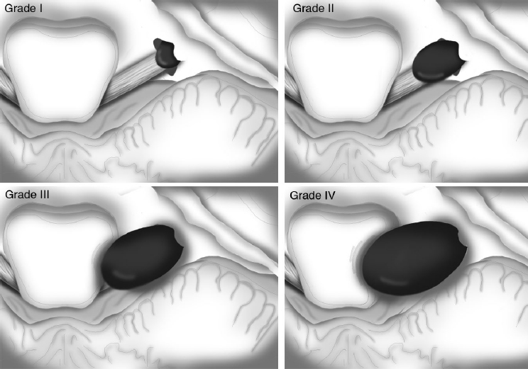
Grading is demonstrated in Table 22.2 and Fig. 22.1.
Table 22.2 Grading of Vestibular Schwannomas
T1 Purely intrameatal |
T2 Intra- and extrameatal |
T3 a Filling the cerebellopontine cistern b Reaching the brainstem |
T4 a Compressing the brainstem b Severely dislocating the brainstem and compressing the fourth ventricle |
Grade 1 Diameter 1–10 mm, intracanalicular |
Grade 2< 20 mm, intracanalicular and intracisternal 2a Tumor does not extend more than 10 mm into the CPA* 2b Tumor extends 11–18 mm into the CPA from the porus acusticus** |
Grade 3< 30 mm, reaching the brainstem |
Grade 4≥ 30 mm, indenting and displacing the brainstem |
Abbreviation: CPA, cerebellopontine angle.
*Measured from the lip of the porus acusticus.
**Leaving 5 to 8 mm between the tumor and the brainstem.
Fig. 22.1 Vestibular schwannomas grading based on the Koos et al classification system. (Adapted from Koos WT, Matula C, Lang J. Color Atlas of Microneurosurgery of Acoustic Neurinomas. New York, Stuttgart: Georg Thieme Verlag; 2002.)
• Grade 4 tumors are associated with a higher incidence of hydrocephalus.
• In fewer than 10% of cases and especially in patients with NF2, the tumor can be purely extracanalicular, growing in the CPA. The rare entity of an extrameatal vestibular schwannoma has been defined as “medial acoustic neuroma,” in which the clinical characteristics are quite different from the other vestibular schwannomas. Medial acoustic neuromas result in preserved hearing and more severe cerebellum, brainstem, and trigeminal impairment.19 Medial acoustic neuromas are often cystic, hypervascular, large, and highly adherent to the brainstem for focal lack of the typically-found duplicated arachnoidal layer.20
Surgical Pathology
It is important to recognize the patterns of displacement of the CPA cranial nerves when approaching the tumor. Review CN VII and VIII anatomy in Chapter 3, see also Fig. 3.3 on page 86.
Facial Nerve
See Table 22.3.
• The nerve can be recognizable in the form of a thin bundle, but in one third of cases it may be splayed out into multiple projections adherent to the capsule (this occurs in two thirds of cases of VS in patients affected by von Recklinghausen’s disease).
Vestibulocochlear Nerve
See Table 22.4.
• In tumors grade 3 and 4, the hearing is lost in 90% of cases. In the few cases when the hearing is functional, such as in cases with a displaced normal nerve, every attempt should be made to preserve it.
Table 22.3 Pattern of Displacement of Cranial Nerve VII9
Pattern | Percent of Cases |
Anterior displacement | 70 |
Superior displacement | 10 |
Posterior course around the tumor | 7 |
Inferior course | 13 |
Table 22.4 Pattern of Displacement of Cranial Nerve VIII9
Type | Pattern | Percent of Cases |
I | The nerve is involved in the tumor, making the separation virtually impossible | 50 |
II | The nerve begins as a bundle from the brainstem, but it splays out in the tumor | 40 |
III | The nerve is spared in its anatomic integrity | 10* |
*It runs medially (18%), laterally (2%), or inferiorly (80%) to the tumor.
• Arterial supply to the tumor: branches from the anterior inferior cerebellar artery (AICA), meningeal branches from internal carotid artery (ICA) and external carotid artery (ECA). In contrast, the rarer variant of hypervascular VSs is supplied by the vertebrobasilar system, presenting several intratumoral arteriovenous shunts.21
Treatment
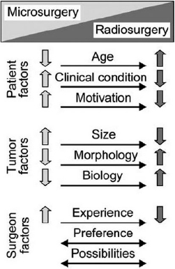
• The type of treatment depends on the size of the tumor, its growth pattern, the neurologic symptoms (cranial nerves or brainstem compression), and patient age and comorbidities (Fig. 22.2).
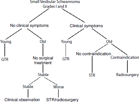
• Small tumors or those found at follow-up that have not shown any growth might be managed conservatively22 (Fig. 22.3), but treatment is suggested when the tumor shows growth over time, there is a worsening of the neurologic symptoms, or new neurologic deficits arise.
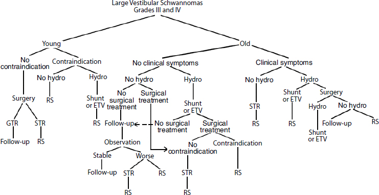
• Gamma Knife radiosurgery is suggested for controlling the growth (with hearing preservation ranging from 64 to 85% and facial nerve preservation in > 95% of cases)11 of tumors smaller than 3 cm or for the treatment of recurrences/residuals of tumors after surgery (see also Chapter 30, page 782). The possible radiosurgical complications, such as hearing loss, facial and trigeminal deficits, hydrocephalus, and potential malignant transformation, must be considered. Other possible complications, such as delayed severe headache, severe facial pain, new motor deficits, or hydrocephalus requiring ventriculoperitoneal shunt, should be considered as well.23 As with surgery, patients with a tumor volume > 5 cm3 have increased rates of complications.24 Regardless of size, Gamma Knife yields better facial nerve results than does open surgery. Surgical resection is still the best treatment modality25 for those healthy patients who are younger than 60 years and have mass effect causing ataxia, severe vertigo or dizziness, brainstem compression, or tumors that are mostly cystic (Figs. 22.3 and 22.4).
Fig. 22.2 Factors to evaluate in determining the treatment for vestibular schwannomas. (From Koos WT, Matula C, Lang J. Color Atlas of Microneurosurgery of Acoustic Neurinomas. New York, Stuttgart: Georg Thieme Verlag; 2002.)
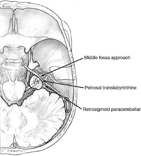
Surgical Approaches (Fig. 22.5)
• Approaches are tailored to the patient, the tumor, and the surgeon’s preferences and experience.
• Surgery is indicated for tumor grades 1 and 2a9 or smaller than 2.5 cm,26 with no functional hearing; a petrosal translabyrinthine approach or retrosigmoid paracerebellar (RSPC) approach may be appropriate.
• For small tumors (grades 1 and 2a) with good hearing, a middle fossa approach (extradural subtemporal approach) or suboccipital transmeatal approach may be appropriate.
• For grade 2, 3, and 4, an RSPC approach may be appropriate.
• A complementary view from an otoneurological surgeon perspective suggests the following algorithm for the selection of the surgical approach22,27: the translabyrinthine approach for tumors > 1.5 cm or for tumors smaller than this size in patients with nonserviceable hearing. Tumors ≤ 1.5 cm in patients with serviceable hearing and tumors < 0.5 cm in patients younger than 65 might be approached via a middle cranial fossa approach; otherwise via a retrosigmoid approach.
• Intraoperative monitoring may be helpful.
• Some surgeons perform a temporary tarsorrhaphy prior to surgery (after anesthesia), and remove it after surgery if there is no facial nerve palsy.
Surgical Pearl
Avoid use of a muscle relaxant for the facial muscles monitoring. Electric stimulation of the facial nerve and evoked muscular responses may be used for prognostication.
Pearl
Transthoracic echocardiography is recommended with the patient in the sitting/semi-sitting position for monitoring air embolism, particularly in patients with a known patent foramen ovale.
• Combined approaches, such as a combined retro- and presigmoid approach or retrosigmoid and middle fossa approaches, may be required for management of giant multicompartmental tumors.
 Surgical Approaches
 Surgical steps of the translabyrinthine approach: see Chapter 14, page 371
 Surgical steps of the retrosigmoid paracerebellar approach: See also Chapter 14, page 385
• All RSPC approaches should expose the sigmoid and transverse sinuses sufficiently so that the view of the surgeon is flush parallel with the petrous face. By drilling the posterior margin of the mastoid (in the so-called extended retrosigmoid approach), the complete exposure of the sigmoid sinus and removal of the mastoid process provides an increase in the angle of work by almost 25 degrees. The mastoid cortex flap can be resected by using chisels instead of being drilled, so that it can be used for reconstruction.28
• Always wax the holes of the mastoid, or pack it by using small pieces of muscle/fat, in order to avoid a CSF leak.

 A cruciate dural opening is also possible but it tends to dry out more than a C-shaped opening (see dural opening options in Chapter 14, Fig. 14.23, page 389).
 Dissect and explore the CPA, and check and coagulate the bridging veins between the cerebellar cortex and the tentorium.
 Preserve the petrosal veins whenever possible.
 Make an interlayer dissection between the two layers of arachnoid: the peripheral layer of the posterior fossa and the layer enveloping the tumor.
Surgical Anatomy Pearl
Check for anatomic variants during dissection (Tables 22.3 and 22.4).
• Check the lateral position of the facial or vestibulocochlear nerves. Perform a stimulation of the posterior and lateral capsule of the tumor to check the position of CN VII, particularly in cases of NF2.
• Check the position of the AICA and its branches, especially the subarcuate artery (going into the subarcuate fossa superiorly to the porus acusticus). Rare variant of AICA: the subarcuate type loops into and out of the dura mater posterior to the internal auditory canal (IAC). In such a case, the dura must be incised around the artery and reflected medially along with the vessel.
 Protect the other cranial nerves by using a gelatin sponge.
 Debulk the tumor’s posterior and central portions (by ultrasonic aspirator, pituitary forceps, and/or a curved cutting instrument).
 Separate the capsule of the tumor from the brainstem, and elevate the inferior pole to find the arachnoid plane separating the tumor from the lower cranial nerves.
 Drill the IAC 180 degrees around. Open the dural envelope in the IAC.
 Dissect the tumor from AICA and CN VIII. Dissect the tumor from the brainstem and cerebellum, starting from the origin of the trigeminal root. Try to remain between the two arachnoid layers, whenever possible. Dissect the lateral pole (Fig. 22.6).
Surgical Anatomy Pearls
• Always check for the anatomic position of the facial nerve. You may use stimulation with low-intensity current (0.1–0.2 mA) to distinguish the facial nerve fascicle from arachnoid bundles and other nerves.
• Facial nerve stimulation at the end of the operation at the brainstem: 0.2 mA, normal function; if no stimulation, with up to a stimulus of 2 mA, the nerve must be inspected. If anatomically interrupted, repair it with microsutures, graft, or hypoglossal-facial anastomosis if the brainstem stump cannot be found (see Chapter 28).29
• Use angled-endoscopes for exploring the CPA and searching for potential remnants, especially in the IAC and around the nerves.
Closure
Closure includes dura mater closure, graft of pericranium, and/or fibrin glue. If craniotomy has been performed, reimplant the bone flap with titanium micro-plates. Use cement or titanium mesh for gaps or in cases of a craniectomy.
 Surgical steps of the middle approach:
 Make the craniotomy as low as possible to reach the middle cranial fossa, otherwise, extend it by drilling the inferior bone margin.
 Elevate the dura over the petrosal surface from posterior to anterior, preserving the greater superficial petrosal nerve.

 Variation: The drilling can be performed according to the Garcia-Ibanez technique by identifying the arcuate eminence and the greater superficial petrosal nerve, and then by drilling between the lines created by these structures for access to the IAC. It is like the Fish technique, and avoids resecting the greater superficial petrosal nerve (GSPN).30 A complete unroofing exposing the geniculate ganglion and the origin of the GSPN may be performed according to the House technique,31 which requires much more time.
 Continue the drilling medial to the cochlea, by using a 2-mm diamond drill, exposing the facial nerve. Open the dura of the IAC, divide the vestibular nerve, and dissect the tumor from its superior aspect. At the end of the resection, fill the space with fat graft.
Vestibular Schwannomas in Neurofibromatosis Type 2
• These tumors can be more challenging because of the multiple surgeries these patients may require and the potential for cumulative neurologic morbidity.32,33 In 40% of cases, these tumors are multilobular, with neurovascular structures passing between the lobules. The tumors can even infiltrate the nerve fibers. Total resection with function preservation is much more demanding, and the morbidity is greater.
• Small tumors should be surgically removed, whereas remnants or recurrences may also be managed by radiotherapy. Surgical removal gives the best outcome, especially in terms of hearing preservation.15 Loss or alteration of waves I and III may be an indication for performing a partial or subtotal resection of the tumor with IAC opening for cochlear nerve decompression.34 Hearing restoration in patients who are already deaf may be attempted with auditory brainstem (midbrain) implants.35–37
• Consider cochlear implants in specific cases (see Chapter 14 on page 383).
• Radiosurgery or fractionated radiotherapy may control the tumor in 81% of patients at 10 years’ follow-up,38,39 with a hearing preservation rate of 33 to 43% (with some deterioration possible also in a delayed fashion over several years).40 Patients with just one side affected could benefit more from surgery, with radiotherapy reserved for older patients or for patients with medical contraindications to major surgery.32,34,41
Outcome
• The overall total removal rate is 70 to 90%.
• The recurrence rate of the tumor (recurring between 1 and 13 years after the first surgery) has been shown to be 0.05% with the enlarged translabyrinthine approach, 0.7% with the retrosigmoid approach, and 1.8% with the middle cranial fossa approach.42
• The retrosigmoid approach is very versatile, most general neurosurgeons are familiar with it.43
Facial Nerve Preservation
Facial nerve preservation depends on the size of the tumor and the preoperative dysfunction, but overall preservation occurs in 82% of cases 1 week after surgery, and 91% of cases 12 to 18 months after surgery.9 The mean preservation is 92 to 96%.21,44 Facial nerve preservation is nearly always possible for tumors smaller than 1 cm.45
• Severe facial nerve palsy (House-Brackmann grade III–VI) may occur in up to 10% of cases for each centimeter in diameter of the tumor.46 It can be temporary (up to 60% of these patients improve over the follow-up). In such cases, tarsorrhaphy is required as soon as possible after surgery. Otherwise, taping the eye for several days and moistening it every 2 hours should be sufficient.
Hearing Preservation
Hearing preservation depends on the size of the tumor as well on the anatomic variant of CN VIII, with an overall rate of 87% in grade 2 tumors18 and 10% in large tumors.
Complications46–48
On a analysis of more than 6,500 patients, the overall complication rate was 28% (26.2% nervous system complications versus 5.1% non–nervous system complications, such as pneumonia, venous thrombosis, acute myocardial infarction, and pulmonary embolism).48
• Overall mortality: < 0.5%
• Facial nerve palsy (see above in the “Facial Nerve Preservation” paragraph)
• Cerebellar contusion/infarction with ataxia: < 1%
• Hematoma: 1%
• Brain infarction/edema (e.g., following occlusion of the vein of Labbé): < 1%
• Brainstem injury: < 1%
• Hydrocephalus: < 4%
• Venous sinus injury/transverse/sigmoid sinus thrombosis: up to 4.5%. It may be related to retraction/manipulation of the sinus during surgery, direct trauma during craniotomy/craniectomy, desiccation of the sinus in long operations, or migration of bone wax.49–51
• CSF leakage: < 7%, pseudomeningocele
1. Reinforcing the wound (more stitches, compressive dressing)
2. Lumbar drainage for 3 to 5 days, draining 5 to 15 cc/h
3. Surgical repair (also waxing the air cells of the mastoid bone)
• Wound infections: 2%
• Meningitis: 1%
• Trigeminal deficit: 2%
• CN IV or VI palsy (diplopia): < 1%
• Lower cranial nerve paralysis (dysphagia, aspiration pneumonia): 0.5%. Arytenoid adduction and thyroplasty for unilateral vocal cord paralysis, nasogastric (NG) tube, and feeding jejunostomy may be required in such cases. See Chapter 8.
• Seizures: up to 7%49
• In cases entailing the harvesting of abdominal fat, a complication rate of 3% for abdominal subcutaneous hematoma has been reported.47
• The postoperative quality of life of patients can be reduced by headache, wound problems, dural irritation, and spasms of the neck muscles related to the surgical position.
Pearl
Patient surveys suggest that the complications with open surgery are worse than those reported by surgeons.
 22.2 Clival, Petroclival, and Petrous Meningiomas
• Posterior fossa meningiomas account for 10% of intracranial meningiomas. Clival and petroclival meningiomas account for 3 to 10% of posterior fossa meningiomas and less than 2% of intracranial meningiomas.1 Meningioma is the second most frequent tumor of the CPA, after vestibular schwannoma (up to 15% of CPA tumors).2,3
• It is rare to see tumors confined to solely one area. Most tumors extend from the clivus to the petrous bone and so are most accurately termed “petroclival.” However, those that take their origin more medially present a greater challenge to the surgeon.
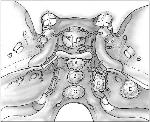
• The petrous face can be divided into three areas: an area medial to Meckel’s cave, a middle area between Meckel’s cave and the IAC, and an area lateral or posterior to the IAC. Tumor localizations are shown in Fig. 22.7. Tumors originating in each of these areas displace cranial nerves and vessels in different ways, and so understanding the origin helps the surgeon to plan the approach and to minimize complications.
• Some of these tumors also extend upward into Meckel’s cave and into the cavernous sinus or the middle fossa. The surgeon must decide which components of the tumor are most responsible for the patient’s symptoms and most amenable to safe surgical extirpation.
• Clival meningiomas have a dural attachment in the midline along of the clivus. Their origin arises medial to the entrance to Meckel’s cave. They displace the brainstem posteriorly.4
• Petroclival meningiomas, or, as they are more appropriately described, medial petrous meningiomas, have a dural attachment at the petroclival junction, medial to the IAC, and lateral to the entrance to Meckel’s cave, posterior to the gasserian ganglion. They displace the brainstem posteriorly and contralaterally.4
• Petrous face meningiomas or CPA meningiomas arise from the dura of the posterior surface of the petrous bone, lateral to the entrance of the trigeminal nerve into Meckel’s cave.5
 Blood Supply of the Tumor
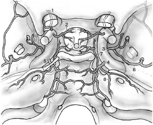
The blood supply is from the clival branches of the meningohypophyseal trunk, branches of the external carotid artery (e.g., ascending pharyngeal), the meningeal artery of the vertebral arteries, or arteries of the vertebral and basilar arteries in the case of pial invasion6 (Fig. 22.8).
 Signs and Symptoms
The signs and symptoms vary based on the position and size of the tumor, with headache and cranial nerve palsies (especially hearing loss, facial numbness/pain, followed by double vision, dysarthria, and dysphagia) occurring most commonly. Tinnitus, ataxia, motor/sensory signs and symptoms, and hydrocephalus are also common.
 Classifications
Clival Meningiomas
Anatomic classification of clivus tumors6 (Fig. 22.7):
• Upper clivus: above the trigeminal nerve root
• Mid-clivus: between the trigeminal root and CN IX
• Lower clivus: from CN IX to the foramen magnum
Petroclival Meningiomas
Petroclival meningiomas are often diagnosed when they have a large or giant size (≥ 4–5 cm in diameter), involving more anatomic compartments.
Petroclival meningiomas are limited to the petroclival region in only 10% of cases. More frequently they involve the Meckel’s cave, and/or the CPA, middle fossa, and foramen magnum.7
Cerebellopontine Angle Meningiomas5,8
Based on the relation of the tumor to the internal acoustic meatus, CPA meningiomas can be:
• postmeatal (± extension into the meatus);
• premeatal (medial and superior extension ± extension into Meckel’s cave, or into the meatus, or medial and inferior extension ± extension into the jugular foramen/meatus, or a combination of these patterns); or
• with a pre- and postmeatal extension.
Petrous face meningiomas can also be classified based on their location relative to the meatus.
 Treatment
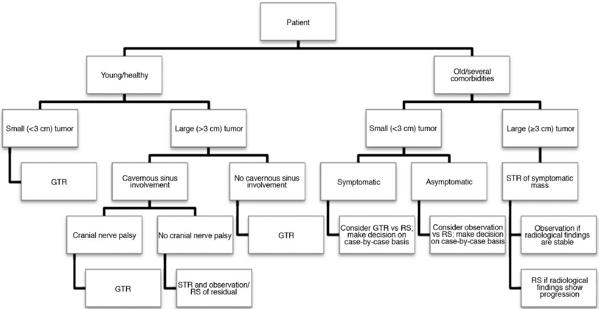
Surgery should be the first-line treatment in young patients and for tumors larger than 3 cm, particularly in patients with significant local mass effect or increased intracranial pressure. Otherwise, careful monitoring, radiosurgery, and fractionated radiation therapy are also options to consider. Fig. 22.9 provides an algorithm of treatment for petroclival meningiomas.9 Surgery is also a very good option for younger patients with smaller tumors and impending neurologic compromise or neurologic symptoms. A total removal in conjunction with excellent long-term control maintains the possibility of preserved function in these patients.10
• Some surgeons have moved to combined-treatment paradigms with more conservative local resections followed by radiosurgery of the remnant. As a result, the total excision rate in some series have dropped recently (from 78–91% in the 1990s to 32–43% in 20071,11–13). Whether this paradigm will achieve better long-lasting control, improved survival, and less overall morbidity remains to be seen.
Pearl
For very small or asymptomatic tumors, observation or radiosurgery alone is also an acceptable option.
 Surgical Approaches
The main goals of surgery are tissue diagnosis; decompression of the brainstem, cerebellum, and cranial nerves; and oncological maximal safe resection. Simpson grade I resection is not always feasible due to the desire to minimize potential neurologic morbidity. Subtotal/partial resection followed by imaging follow-up and/or radiotherapy often can be considered adequate for a prolonged good clinical outcome.
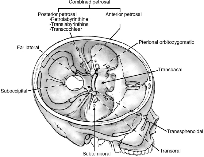
• A number of surgical approaches are possible for these tumors (Table 22.5 and Fig. 22.10). The approach should be tailored to the patient and the actual extent of the tumor. For those tumors that extend through the tentorial incisura into the middle fossa, a combined transtentorial infra-supratentorial approach with or without a posterior or anterior petrosectomy is helpful. For those lesions that are confined within the posterior fossa, a laterally placed posterior fossa craniotomy flush with the face of the petrous bone with or without a retrolabyrinthine posterior petrosectomy often provides a wide and efficacious corridor. The extended transtemporal exposures (extended retrolabyrinthine, transcochlear) approaches are also excellent ways to access these lesions. Occasionally, an isolated lesion presents purely anteriorly along the clivus and an extended transsphenoidal approach may be appropriate. For posterolateral approaches, see Chapter 14, page 384.
Table 22.5 Approaches Used for Clival, Petroclival, and Cerebellopontine Angle (CPA) Meningiomas
Approach | Variant/Extension |
Retrosigmoid paracerebellar | • Presigmoid exposure* • ±Partial/total labyrinthectomy • ±Transcochlear/total petrosectomy |
Frontotemporal-orbitozygomatic | • Alone or in combination with other approaches • Transcavernous approach |
Posterior petrosal | • Partial/total labyrinthectomy • Transcochlear/total petrosectomy |
Subtemporal transtentorial | • Extended middle fossa (anterior petrosectomy) • ± Retrosigmoid paracerebellar • ± Transtentorial |
Far lateral/extreme lateral | • Transcondylar • Retrocondylar |
Extended transsphenoidal | • Transclival • Posterior clinoidectomy, pituitary transposition |
Transbasal |
|
Transoral |
|
Suboccipital |
|
Note: See text for indications. Several combinations of these approaches can be performed.
 Surgical Positions
The patient is placed in the supine position with a large roll used to elevate the ipsilateral shoulder. The head is fixed in pins and turned 70 degrees to the contralateral side. A lateral position or park-bench position can also be used. Some surgeons prefer placing the patient in the sitting position. The extended transtemporal procedures are done with the patient in the supine position with the head turned, with or without a shoulder roll. All pressure points should be carefully padded.
The space between the shoulder and the ear should be maximized by a number of maneuvers: tilting the ipsilateral shoulder inferiorly and anteriorly away from the ear, padding it, and taping it there; and tilting the vertex of the head down toward the floor. This can be facilitated by allowing the dependent arm, in the lateral or park-bench position, to hang below the level of the bed on a well-padded arm board. Once the patient is positioned and carefully secured in place, the operating table should be tilted laterally and in the Trendelenburg/anti-Trendelenburg position before starting the operation to ensure that the patient is stable and will not move during bed repositioning intraoperatively and that any lines or the endotracheal tube will not be at risk of disruption during movements of the bed. Prepare and drape the patient’s abdomen or the lateral thigh for fascia lata and fat graft, which are later required for closure.
• Intraoperative monitoring is useful for the dissection of cranial nerves.
• Note that all retrosigmoid paracerebellar approaches should carry the craniotomy far lateral enough so as to clearly expose the transverse and sigmoid sinuses. The CSF should also be drained from the cisterna magna routinely in all cases for posterior fossa meningioma prior to the main opening of the dura. This usually requires drilling away some of the mastoid. This technique brings the surgeon parallel with the petrous face and avoids the need to retract the cerebellum.
• With appropriate brain relaxation, an approach flush parallel with the petrous face, and extensive initial internal debulking of the mass, the RSPC posterior fossa craniotomy approach can access most of the lesions without brain retraction.
• The RSPC approach can be combined with a variety of extensions medial to the sigmoid sinus—the so-called presigmoid exposure. The smallest of these involves removing the bone in Trautmann’s triangle medial and anterior to the sigmoid sinus by a simple mastoidectomy that preserves the labyrinth and otic capsule. More extensive bone removal can include removal of a part of the labyrinth (partial labyrinthectomy) or all of it (total labyrinthectomy, or doing a total labyrinthectomy with removal of the cochlea and anterior medial petrous apex (transcochlear and total petrosectomy).
• For small or medium-size, laterally located tumors of the petrous face or mid-clivus or larger tumors of the lateral petrous face or CPA: retrosigmoid paracerebellar approach or a presigmoid petrosal approach without labyrinthectomy (retrolabyrinthine petrosal approach).
• For small or medium-size, centrally located tumors of the upper or mid-clivus (when hearing is unaffected): retrosigmoid paracerebellar approach, posterior petrosal presigmoid approach, or posterior petrosal partial labyrinthectomy approach.
• For small or medium-size, centrally placed tumors of the upper or mid-clivus (when hearing is not preserved): retrosigmoid paracerebellar approach or translabyrinthine petrosal approach, providing additional exposure to the IAC.
• For retro-meatal petrous face tumors: retrosigmoid paracerebellar approach.
• For tumors located at the lower clivus: retrosigmoid paracerebellar approach or extreme lateral transcondylar approach if the surgical corridor is small.14
• For lateral tumors of the petrous ridge or tumors in the CPA: retrosigmoid paracerebellar approach15 or retrosigmoid suprameatal approach. Rarely, a retrosigmoid craniotomy and intradural resection of the petrous apex with drilling and removal of the suprameatal tubercle can be used to manage CPA meningiomas when there is difficulty reaching the suprameatal components.
• Combined approaches may be required for tumors with extension above and below the dorsum sellae.
• The frontotemporal-orbitozygomatic (FTOZ) approach may be performed for tumors extending superiorly to the dorsum sellae, alone or alongside other approaches, as indicated by the size and localization of the tumor.
• Clival/petroclival meningiomas can also be approached by means of the endoscopic extended endonasal transclival approach.16 An endoscopic endonasal transcavernous posterior clinoidectomy with interdural pituitary transposition has been suggested as well.17
• Further approaches to the clival and petroclival tumors: transbasal, transoral, subtemporal, suboccipital, and far lateral.
• Retrosigmoid intradural suprameatal and retrosigmoid transtentorial approaches provide adequate exposure to the petroclival regions.
• The anterior upper third of the posterior fossa can also be reached by a transcavernous approach for the treatment of selected petroclival meningiomas, trigeminal schwannomas, and clival chordomas, but it is rarely used.18
 Posterior Petrosal Approach Technique
 Incision. A number of incisions are acceptable. The most common is likely a C-shaped incision along or below the superior temporal line to reach the retroauricular region. For orbitozygomatic (OZ) combined approaches, the incision should come down anterior to the tragus and an extension to the frontal region is added to provide access to the orbital margin (see Chapter 14, Fig. 14.22, page 388).
 Elevate the pericranium and the temporal muscle, and reflect the muscle anteriorly.
 The sternocleidomastoid muscle can be reflected anteriorly with the trapezius, semispinalis capitis, and splenius capitis muscles inferiorly and posteriorly, for optimal exposure and reconstruction at the end of the operation.
• For all but the smallest tumors, all the approaches with presigmoid exposures are combined with exposure of the dura posterior to the sigmoid sinus via a posterior fossa craniotomy or craniectomy.
• Many authors call the posterior petrosal approaches simply the “petrosal approaches.” Here the term posterior petrosal is used to distinguish these approaches from the anterior petrosal approach popularized by Kawase et al,19 which are performed from a middle fossa exposure. These petrosal approaches include exposure of the dura anterior and medial to the sigmoid sinus, and then varying amounts of removal of the otic capsule (labyrinth, cochlea) and ultimately the petrous apex in a total petrosectomy.20
 Expose the temporal, retrosigmoid, and mastoid areas, as well as the root of the zygoma.
 Identify the position of the venous sinuses.
 Craniectomy and craniotomy (using one or more bur holes) are two viable alternatives. Bur holes can be placed on the transverse sinus, taking care not to injure the mastoid emissary vein.

 Variation: Perform retrosigmoid craniotomy, mastoidectomy, and/or further exposures according to the planned approach.
Surgical Anatomy Pearl
Extension of the craniotomy: it should be larger (at least ~ 1 cm) anteriorly and posteriorly to the extent of the tumor (as seen in preoperative neuronavigation).
 Retrolabyrinthine presigmoid approach:
 Perform mastoidectomy and partially unroof the sigmoid sinus, semicircular canals, vestibular aqueduct, jugular bulb, and mastoid segment of the facial nerve in order to leave these structures in a partial bone “shell” for protection. The sinodural angle must be exposed.

 Variation: Partial labyrinthectomy/petrous apicectomy:
• Perform a complete cortical mastoidectomy; see the position of the semicircular canals (blue line).
• Prior to opening the canals, wax them to avoid loss of endolymphatic fluid and hearing loss. Despite taking this step, however, the majority of patients will still lose hearing.
• Perform a partial labyrinthectomy for the petrous apicectomy, between the ampulla of the superior semicircular canal and the entrance of the vestibular aqueduct into the petrous dura.21
• Skeletonize the superior wall of the IAC.
 After the mastoidectomy, resect the semicircular canals and open the vestibule.
Surgical Anatomy Pearl
Exposure of the presigmoid dura should be maximized while also removing as few temporal bone structures as possible.
 Skeletonize the facial nerve from its genu to the stylomastoid foramen.
 Resect the bone of the IAC.
• At the end of these surgical steps, the visible walls in the surgical corridors are the sigmoid sinus posteriorly, the tegmen dura superiorly, the facial nerve anteriorly, and the jugular bulb inferiorly (see Chapter 14, Fig. 14.20, page 374).

 Variation with total petrosectomy: reserved for giant midline tumors. It can be useful in those cases with prior surgery or radiation, bilateral extension of the tumor, or extensive vascularization. Often the procedure can be multistaged.
 The exposure is similar to that of the other petrosal approaches, but transecting and over-sewing the external auditory canal is required.22

 Variation: Dissect the temporomandibular joint capsule from the glenoid fossa.
 Perform a labyrinthectomy with exposure of the facial nerve.
 For lesions with significant supratentorial extension, a temporal craniotomy with or without a zygomatic osteotomy including the condylar fossa, with further resection of the condyle and neck of the mandible, may be required if access to the horizontal petrous segment of the ICA is needed.22
 Identify and resect the greater superficial petrosal nerve and the middle meningeal artery.
 Expose, pack with autologous fat, and suture the cartilaginous eustachian tube.22

 Variation: Unroof the petrous ICA. Divide the fibrocartilaginous ring around the cervical carotid at its entrance into the skull base for anterior mobilization of the ICA.

 Variation: Remove the cochlea and bone medial to the mastoid segment and posteriorly mobilize the facial nerve.
 Perform a medial petrous apex and lateral clivus resection.

 In multistaged operations, at this point, the first step could be stopped, and the facial nerve should be covered with Gelfoam before closure and reopening for tumor resection in the second stage of the operation.
Begin with the cisterna magna or in the presigmoid region, from the inferior part of the sigmoid sinus, parallel to the jugular bulb.
 The presigmoid incision should come up to the superior petrosal sinus. The temporal dura should be opened parallel to the floor of the middle fossa.
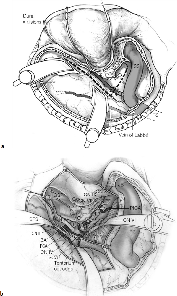
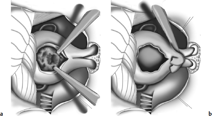
 When the incisions join the superior petrosal sinus, the latter should be suture ligated and divided (Fig. 22.11a).
 Next, divide the tentorium.
 Put sutures on the dural edges for the augmentation of the space.
 Cauterize the superior petrosal veins, whenever required.
Surgical Anatomy Pearl
Open the basal cisterns for CSF drainage.
Surgical Anatomy Pearl
Be mindful of the vein of Labbé and ensure that it sustains no damage.
 Open Meckel’s cave to mobilize and visualize the trigeminal root. Open the arachnoid between the nerves.
 Start tumor debulking between CNs IV and V and CNs V and VII to VIII, to minimize the manipulation of the nerve complexes.6
 Proceed toward the base and inferior pole of the tumor and identify CN VI, disconnecting and coagulating the petroclival dura.
 Dissect the other nerves and the basilar artery and its branches.
 Resect any potential intracavernous extension of the tumor (although for intracavernous tumor components it is better to combine this approach with a subtemporal approach and OZ osteotomy).
Surgical Anatomy Pearl
Preserve the arachnoid plane for the brainstem decompression.
Surgical Anatomy Pearls
• Be mindful of the superior cerebellar artery (SCA) and CN IV and ensure that they sustain no damage (Fig. 22.11b).
• Tentorial detachment is an important requirement in combined petrosal approaches in order to provide access to medium-to-large petroclival tumors and to facilitate their resection. The preoperative understanding of the anatomic relationships between the tentorium and the temporal bridging veins as well as with CNs IV to VI is of paramount importance.23
• A petrosal approach with tentorial resection generally requires sectioning of the superior petrosal sinus, although the dura may be opened more anteriorly to it, in order to preserve it, by means of further drilling of the medial wall of the petrous bone, to reach anteriorly up to the suprameatal area. The separation of the tentorium from Meckel’s cave in fact enables a connection between the middle and posterior cranial fossa with a partial opening of the tentorium and preservation of the superior petrosal sinus.24
Wherever possible, perform primary dural closure. Pack the eustachian tube well. However, the presigmoid exposure often requires duraplasty with autologous tissue such as fascia lata and fat graft.
 Fill the presigmoid space or the space of the total petrosectomy with autologous fat.
 Reinforce it with fibrin sealants.

 Some surgeons add a titanium mesh cranioplasty, miniplates, or hydroxyapatite cement to improve cosmesis.

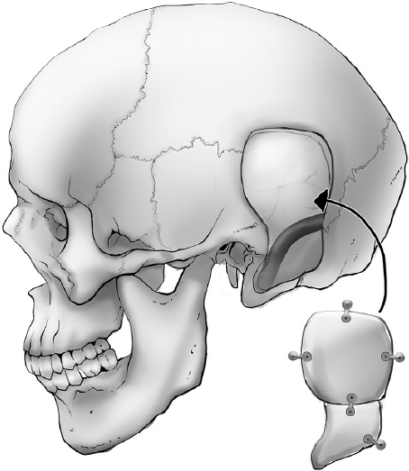
 Some surgeons use the “split technique” to cover the mastoid defect (Fig. 22.12).
Fig. 22.12 Split technique. The temporal bone flap can be split with a reciprocating saw, providing a split calvarial bone to cover the mastoidectomy defect. (Adapted from Bogaev C, Sekhar LN. Petroclival meningiomas. In: Sekhar LN, Fessler RG, eds. Atlas of Neurosurgical Techniques. New York: Thieme; 2006:695–710.)
 Suture the temporal and retroauricular muscles.

 Variation: Microplate-Bridge technique: a fascial graft fixed with a long micro-titanium plate over the presigmoid-subtemporal defect may help prevent CSF leak.25
 Outcome
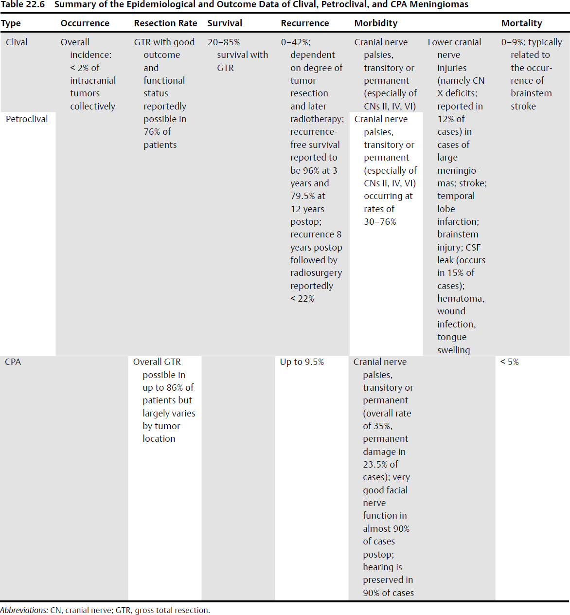
Surgical radicality and morbidity are related to the different tumor types (Table 22.6).
Clival and Petroclival Meningioma
Gross total resection: 20 to 85% survival,7,26–29 dependent on tumor size, consistency, invasion of the cavernous sinus, and cranial nerve involvement.30 Skull base approaches enable a Simpson grade I or II resection (total removal) with good outcome and functional status in 76% of patients.31 The recurrence rate is dependent on the degree of resection, as well as on later radiotherapy. Recurrence-free survival has been reported up to 96% at 3 years and 79.5% at 12 years.1 As a result, the recurrence rate ranges from 0 to 42%.32 Recurrence at 8 years after surgery followed by radiosurgery has been reported as less than 22%.1,7,26,32
Cerebellopontine Angle Meningioma
Overall gross total removal (Simpson grade I and II) is possible in up to 86% of patients,33 although the degree of resection correlates with the tumor topographic location. The degree of resection is highest in the suprameatal and retromeatal subtypes, and lowest in the premeatal and inframeatal types.8
Mortality
The mortality rate in clival-petroclival meningioma is 0 to 9%, largely caused by brainstem stroke,7,29,30,32 and below 5% in CPA meningiomas.33
Morbidity
Morbidity entails cranial nerve palsies (new or worsening of preexisting conditions), which can be transitory or permanent, especially of CNs II, IV, and VI in clival and petroclival meningiomas.29,30 Cranial nerve deficits occur at a rate of 30 to 76%29 in petroclival meningioma surgery, according to the different published series. A preoperative low Karnofsky performance score, age ≥ 60, brainstem edema, absence of the arachnoidal plane, vessel/CNs encasement, and hard consistency of the tumor are among the factors contributing to a worse outcome.7,28
• CPA meningiomas: The overall CN deficit rate is approximately 35% (permanent in 23.5% of cases).34 Some series report very good facial nerve function (House-Brackmann grades 1 to 2) in almost 90% of cases of CPA meningiomas after resection, especially in the retromeatal types,33 with a preservation of hearing in 90% of cases.8
Pearl
CPA meningiomas have a recurrence rate up to 9.5%,3,35,36 thereby providing impetus for regular radiological follow-up.33
• Lower cranial nerve injuries can occur in large meningiomas.37–39 CN X deficit has been reported in up to 12% of cases.34
• Stroke, temporal lobe infarction, brainstem injury, and CSF leak (occurrence rate up to 15%) are also possible, especially in combined approaches. Other reported complications include hematoma, wound infection, and tongue swelling.
Pearl
Petroclival meningioma surgery significantly affects patients’ health and quality of life for several years, suggesting that patients’ psychosocial support and rehabilitative measures should also be improved.1,39
 22.3 Epidermoid and Dermoid Tumors
• These tumors account for 1% of intracranial tumors, with an epidermoid/dermoid ratio of 4:1, and account for 7% of CPA tumors (they can also arise in the parasellar region or other locations). In the posterior fossa, another typical localization is the fourth ventricle (more common for dermoids).
• Epidermoid tumors at the CPA are also called cholesteatomas, although this definition is used more for middle ear lesions, with chronic middle ear infections giving rise to entrapped epithelium, forming a kind of pocket that may or may not extend into the posterior fossa.
Surgical Anatomy Pearl
Typically, epidermoids occupy the lateral position (CPA) and the dermoids the midline (Table 22.7). Other skull base locations include the petrous apex.
Table 22.7 Comparison of Epidermoids and Dermoids
Feature | Epidermoid | Dermoid |
Frequency | 0.5–1.5% of brain tumors | 0.3% of brain tumors |
Lining | Stratified squamous epithelium | Also include dermal appendage organs (hair follicles and sebaceous glands) |
Contents | Keratin, cellular debris, and cholesterol; occasional hair | Same as epidermoids + hair and sebum |
Location | More common laterally (e.g., CPA) | More commonly near midline |
Radiology | High signal on DW-MRI, for differentiation from CSF | Variable intensities and calcium from dental elements |
Associated anomalies | Tend to be isolated lesions | Associated with other congenital anomalies in up to 50% cases |
Meningitis | May have recurrent aseptic meningitis (including Mollaret’s meningitis) | May have repeated bouts of bacterial meningitis |
Abbreviations: CPA, cerebellopontine angle; CSF, cerebrospinal fluid; DW-MRI, diffusion-weighted magnetic resonance imaging.
Source: Adapted from Greenberg MS. Handbook of Neurosurgery, 7th ed. New York: Thieme; 2010.
• Very few cases of malignant transformation of an epidermoid cyst into carcinomas have been reported.1
 Pathology
Epidermoids or so-called pearly tumors (because of their shiny white pearl–like appearance) grow due to the accumulation of keratin and cholesterol, expanding and filling the subarachnoid spaces. Dermoids are made up of stratified squamous epithelium, containing dermal elements, including hair, sebaceous glands, or even teeth. See also Chapter 6, page 153.
 Radiology
Epidermoids show high signal on diffusion-weighted (DW) MRI, for differentiation from CSF; dermoids may show variable intensities and calcium from dental elements. See also Chapter 5, page 126.
 Signs and Symptoms
The signs and symptoms depend on the location of the lesion. In the CPA, cranial neuropathies are noted: CN V, isolated trigeminal neuralgia; CN VII, hemifacial spasm; CN VIII, sudden hearing loss. Cranial nerve palsies are related to the tumor size and position, with possible involvement of the lower cranial nerves as well, and hydrocephalus/intracranial hypertension. A rare cause of aseptic meningitis (Mollaret’s meningitis) is related to spilling of intracystic components within the CSF.2,3 These patients present with a sudden severe headache reminiscent of a subarachnoid hemorrhage, but MRI shows fat density globules in the subarachnoid space. Patients usually recover over days to weeks.
 Treatment
Conservative treatment with clinical and radiological follow-up is an option in asymptomatic/debilitated patients, but surgery remains the gold standard of treatment, with no role for radiotherapy. Tumors are very soft and avascular, and can be debulked piecemeal.
 Surgical Approaches for CPA Epidermoids
The retrosigmoid approach is most commonly used for the resection of CPA epidermoids. Transpetrous approaches are generally reserved for specific cases of petrous apex epidermoids, or large cholesteatomas involving the middle ear and temporal bone.
Surgical Anatomy Pearls
• The capsule of the tumor should be carefully dissected and removed, except in cases when it is too tightly adhered to cranial nerves, blood vessels, and/or the brainstem. Blood vessels and cranial nerves are generally engulfed, and therefore the resection has to be done carefully, in piecemeal, and often incompletely to avoid vascular damage and/or neurological dysfunction. An ultrasonic aspirator is rarely needed but may also speed up tumor debulking.
• Try to avoid spilling of intracystic contents within the CSF, so as to preclude chemical meningitis or hydrocephalus. To contain any spillage of contents, protect the surrounding structures with cottonoids. Use copious saline irrigation of the surgical field. Some authors advise utilizing hydrocortisone or perioperative steroids but without evidence.4
• Intraoperative cranial nerves monitoring is helpful.
• Angled endoscopes can help in the microsurgical approach (or in pure endoscopic approaches) to ensure a greater degree of tumor resection by looking around the vessels and cranial nerves in areas that cannot be otherwise observed.5–7
 Outcome
Resection can be total or subtotal and in cases in which the cyst or some component of the tumor is stuck to the brainstem/nerves/vessels, these adherent sections are left in order to avoid/decrease the risk of morbidity.8 Symptoms such as trigeminal neuralgia and headache are often relieved for years due to the effective debulking. MRI every year or every other year with diffusion sequences is used to monitor possible regrowth of the lesion, which may occur even beyond a decade.9 Symptomatic recurrences, occurring after subtotal removal, may require further surgery after an extended period of time because of their slow growth rate. Mortality is rare and recurrences are related to the amount of tumor remnant from the initial surgery.
 22.4 Chordoma and Chondrosarcoma
 Epidemiology
Chordoma and chondrosarcoma each account for 0.15% of intracranial tumors. Chordomas occur at an incidence rate of 0.5 to 8 cases per million persons per year,1–4 32% of which have cranial localization.3 In general, chordomas are located at the midline of the skull base (i.e., clival chordoma), whereas chondrosarcoma are paramedian (at the petrosphenoclival synchondrosis) (Table 22.8).
 Signs and Symptoms
These tumors can give rise to diplopia, proptosis, facial pain, headache, retro-orbital pain/pressure, cranial nerve palsy (above all in CN VI), visual loss, otalgia, nasal obstruction, epistaxis, dysphonia, and dysphagia, depending on the tumor location and invasion of other anatomic structures.
• Occipitocervical pain may occur in cases of instability due to invasion of the occipitocervical joint.
• In cases of brainstem compression, long tract signs may be present.
• Signs of increased intracranial pressure are possible.
Table 22.8 Characteristics of Chordomas and Chondrosarcoma
Characteristic | Chordoma | Chondrosarcoma |
Percent of brain tumors | 0.15% | 0.15% |
Origin | Notochordal remnants | Embryonal mesenchymal remnants |
Skull base location | Midline paramedian | Paramedian |
Age range (years) Median age | 6–78 45 | 25–57 40 |
Male/female ratio | 1.5:1 | 3.7:1 |
Pathological types | Chondroid Nonchondroid Dedifferentiated | Classical (grade I, II, III) Mesenchymal Dedifferentiated |
Histopathological markers | EMA, cytokeratin, and brachyury + | EMA, cytokeratin, and brachyury − |
MRI marker | Increase in ADC values | Decrease in ADC values |
Abbreviations: ADC, apparent diffusion coefficient; EMA, epithelial membrane antigen.
Source: Adapted from Rostomily RC, Sekhar LN, Elahi F. Chordomas and chondrosarcomas. In: Atlas of Neurosurgical Techniques. Brain. Sekhar, L.N.; Fessler, R.G. ed. New York, Stuttgart: Thieme; 2006:778–810.
 Gross Pathology
Chordoma and chondrosarcoma are lytic, expansile, and locally invasive into surrounding structures (clivus, cavernous sinuses, petrous bone, craniocervical junction). The extent of their invasion influences the surgical approach. The tumors are generally extradural and are also not very vascularized and generally quite soft (cartilaginous, similar to vertebral disks) or gelatinous. Calcifications make the resection more difficult.5,6
• Chordomas have a slow and progressive growth and tend to be very invasive locally. Some patients die due to local tumor effects, and not as a result of metastases.
• Chordomas are much more aggressive than chondrosarcomas of the skull base.7–10
• Chordomas and chondrosarcomas require a multidisciplinary team approach for increasing the survival time, improving the quality of life, and lowering the morbidity.11
 Radiology
For lytic lesions, a CT scan is performed to demonstrate skull base erosions and tumoral calcifications.
On MRI, both tumors are iso-hypointense on T1- and hyperintense on T2-weighted images. Diffusion-weighted MRI may be useful in differentiating chordoma from chondrosarcoma, with chondrosarcoma having higher apparent diffusion coefficient (ADC) values.12 See also Chapter 5.
• Chordomas may develop anteriorly, toward the sphenoid sinus; laterally, into the CPA; inferiorly toward the foramen magnum and spinal canal, with invasion of the retropharyngeal space also possible.
• Differential diagnosis with ecchordosis physaliphora (benign notochordal remnant at the dorsum sellae, generally asymptomatic, but can still cause headache, diplopia, pain). In ecchordosis, there is no gadolinium enhancement.13
 Other Lesions Found in the Clivus Area
Other lesions include meningioma, metastasis, plasmacytoma, lymphoma, neuroenteric cyst, retroclival components of craniopharyngiomas and pituitary adenomas, mucocele of the sphenoid sinus, nasopharyngeal carcinoma and rhabdomyosarcoma, and chondromyxoid fibroma.
• The preoperative radiological workup includes CTA/MRA for assessment of the tumoral and peritumoral vascularization.
 Treatment
Surgery is the main treatment for these lesions and is performed for tissue diagnosis, tumor debulking, and maximal safe oncological resection. Almost all cases of chordoma are followed by high-dose radiotherapy because of the radioresistance of the lesion. Despite aggressive surgical resection and radiation, 20% of chordoma cases recur within 1 year.14 Debate exists over whether low-grade chondrosarcoma that is fully resected requires radiation, as the behavior of chondrosarcoma can be quite variable.
• Survival is related to the degree of resection: 5-year survival rate: 90% for chondrosarcoma and 65% for chordoma after gross total resection, with percentages decreasing in cases of partial resection.
 Radiotherapy
See Chapter 30, pages 777 and 779.
 Surgical Approaches (Table 22.9)
Given the size of the tumor, patient signs and symptoms, and general condition of the patient, the surgical options are biopsy followed by observation or radiation, or surgical resection with or without radiation. In symptomatic patients, the best treatment is surgery (as extensive as possible) followed by radiation. Radical resection of chordomas and chondrosarcoma improves the long-term prognosis.15–17
• Eroded/infiltrated bones as well as infiltrated dura should be resected as well. Intratumoral calcifications may occur, especially in chondrosarcoma, and should be drilled and removed.
• Intraoperative monitoring of lower cranial nerves can be useful in surgery. Preoperative balloon test occlusion of the carotid or vertebral artery may be useful if surgical sacrifice might be needed, but the rate of false negatives is high and the test itself entails a risk of stroke.
• The approach depends on the exact location and extent of the tumor. The approaches can be anterior (e.g., transfacial, subfrontal, endoscopic trans nasal/transoral, extended transsphenoidal or combined), anterolateral (e.g., FTOZ), lateral (e.g., subtemporal with anterior petrosectomy), posterolateral (e.g., retrosigmoid, translabyrinthine), or inferolateral (e.g., far-lateral approaches).
Table 22.9 Approaches Used for Chordoma and Chondrosarcoma
Approach | Variant/Extension |
Transfacial | • Facial translocation • Le Fort I transmaxillary transpterygoid approach • Transoral ± extended transsphenoidal • Cervical retropharyngeal |
Extended subfrontal |
|
Frontotemporalorbitozygomatic | • Transcavernous • ± Subtemporal |
Subtemporal | • Extended middle fossa (anterior petrosectomy) • ± Retrosigmoid paracerebellar • ± Transtentorial |
Far lateral/extreme lateral | • Transcondylar • Retrocondylar |
Extended transsphenoidal | • Transclival • Posterior clinoidectomy, pituitary transposition |
Note: See text for indications. Several combinations of these approaches can be performed.
1. Transfacial approaches: include facial translocation (and all its variants), Le Fort I transmaxillary transpterygoid approach,18 transoral or extended transsphenoidal, and high anterior cervical retropharyngeal approaches for midline-paramedial lesions (see Chapter 15). The use of retractors in natural cavities (nasal, paranasal, and mouth) widens the surgical corridors and provides direct ventral access to the target, without any brain manipulation. Postoperative facial swelling is generally tolerated by the patient, but there are risks of speech, swallowing, or airway dysfunction, cosmetic problems (with accompanying psychological problems and worse quality of life), and infections.
2. Extended subfrontal approaches: provide access to the midline clivus, medial cavernous sinus, petrous apex, occipital condyles, and foramen magnum.19 Tumoral components too lateral or at the level of the dorsum sellae cannot be reached. For the technical steps, see page 343.
3. FTOZ approach with transcavernous route: used for management of intracavernous tumor components. Combined with a subtemporal approach, the upper clivus and petrous apex are accessible.
4. Subtemporal transpetrous approach: provides access to tumor components in the middle cranial fossa, upper clivus, and cavernous sinus.
5. Subtemporal-infratemporal approach: extends more inferiorly than the OZ approach in order to reach the horizontal segment of the petrous ICA,20 the parapharyngeal space, orbit, and paranasal sinuses in the event of their involvement. The OZ approach would include the glenoid fossa. This approach also may be used in cases where ICA bypass is deemed necessary.
6. Transpetrosal approach: Determining the tumor extension (upper, middle, or lower clivus, and lateral extension) enables the surgeon to choose the type of petrosectomy (such as Kawase’s approach in the subtemporal craniotomy, posterior petrosectomy, translabyrinthine, or transcochlear approach). For the technical steps, see Chapter 14 and page 573.
7. Lateral transcondylar approach: used for managing tumors in the upper ventral cervical spine, lower clivus, foramen magnum, and occipital condyles.21 This approach requires extraperiosteal management of the vertebral artery, especially for tumors extending laterally to it.15 In some cases, its transposition is necessary, providing wider exposure of the C0-C1 and C1-C2 joints. This approach and any posterior fossa approach can be complicated by cerebellar damage, vertebral artery injury, lower cranial nerve injury, or occipitocervical instability (occurring when > 50% of the condyle is resected).

 The far lateral approach (dorsal transcranial transcondylar) may be associated with the ventral endoscopic endonasal transcondylar (far medial approach) for resection of extensive lesions involving both ventromedial and dorsolateral compartments.22
Surgical Anatomy Pearl
If more than 50% of the condyle is resected, occipital cervical fusion is usually advised.
Stay updated, free articles. Join our Telegram channel

Full access? Get Clinical Tree


 Identify the edge of the petrous ridge, and drill Kawase’s area to the superior semicircular canal and to the IAC.
 Management options:

 Variation with translabyrinthine approach: faster than the partial labyrinthectomy/petrous apicectomy approach, it also provides more exposure of the IAC.
