♦ Preoperative
Evaluation
- Detailed CN examination
- Computed tomography of temporal bone
- Assess anatomic relationship between bone and lesion
- Assess behavior of lesion with respect to bone (i.e., osteodestruction, hyperostosis, etc.)
- Assess anatomy of middle ear
- Assess anatomic relationship between bone and lesion
- Magnetic resonance imaging with and without gadolinium
- Assess soft tissue characteristics, including lesion, brain, vessels, CNs
- Consider magnetic resonance venogram to assess sinus dominance/patency
- Consider cerebral angiography for preoperative embolization of hypervascular lesions, assessment of temporal and posterior fossa venous anatomy, vertebrobasilar anatomy
Special Equipment
- Headlight/loupes
- Mayfield head clamp
- High-speed drill
- Microscope
- Neurophysiologic monitoring: somatosensory evoked potential, motor evoked potentials, brain stem auditory evoked responses, and facial electromyography
- Facial nerve stimulator (Kartush [Medtronic, Minneapolis, MN])
- Ultrasonic aspirator
- Facial nerve stimulator (Kartush [Medtronic, Minneapolis, MN])
Anesthetic Issues
- Allow for neurophysiologic monitoring
- Arterial line
- Central venous access
- Sequential compression devices/thromboembolism deterrent stockings
- Dexamethasone 10 mg intravenous (IV) for intradural lesions
- Mannitol 0.5 to 1 g/kg IV bolus for intradural lesions
- Antibiotics
- Normocapnia
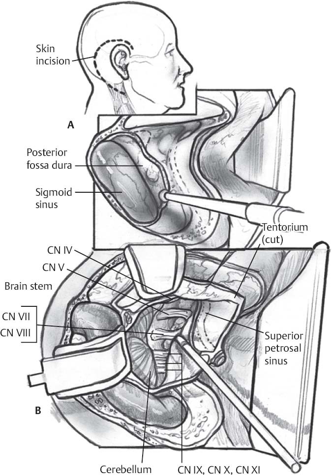
♦ Intraoperative (Fig. 13.1)
Positioning
- Supine with head turned ~45 to 60 degrees and tilted toward dependent shoulder
- Too much rotation may compromise venous drainage
- With larger patients, a shoulder roll or lateral positioning may be necessary for adequate neck rotation
- Mayfield head pins: single pin on ipsilateral frontal bone lateral and posterior to supraorbital nerve, double pin on contralateral occipital bone
- Secure patients firmly with straps to allow intraoperative rotation
- Tape ipsilateral shoulder to foot of table, avoiding excess tension on brachial plexus
- All pressure points must be well padded to avoid decubiti
- Prepare and drape abdomen for free fat graft
Incision
- C-shaped incision beginning anteriorly at the level of the root of the zygoma, two fingerbreadths cranial to the pinna, extending posteriorly in a line parallel to the transverse sinus, then caudally two fingerbreadths posterior to the mastoid process, then anterolaterally along the border of the sternocleidomastoid muscle
Soft Tissue
- Incision is made through galea with care taken to preserve the temporalis and nuchal muscular fascia and pericranium. The flap is reflected anteriorly to the level of the external auditory canal and held in place with suture or self retaining hooks.
- The common musculofascial cuff of the temporalis and nuchal muscles is divided, and the muscles are reflected cranially and caudally, respectively.
Only gold members can continue reading. Log In or Register to continue


