Radiologic Assessment of All Unfused Lumbar Segments After Instrumented Posterior Spinal Fusion
Ferran Pellisé
Alejandro Hernández
Accelerated segment degeneration (ASD) adjacent to a lumbar fusion (LF) has been reported in several papers (1,1,3). However, most of the evidence is derived from series that have not accounted for the natural history of degenerative changes developing at the unfused levels (4). Several experimental studies have suggested an important role for biomechanical changes as the origin or cause of ASD (4,5,6,7). Fusion, especially posterior instrumented, produces increased stress at the adjacent segment, which might be the cause of this syndrome; the stress would be affected by the length, location, and stiffness of the fusion mass (4,5,6,8,9,10). However, one should be cautious when considering the value of much in vitro data because the in vivo situation might be much more complex than is reproduced in vitro (11). Some investigators believe that degenerative changes appearing in the nonoperated segments after fusion may be predetermined by factors other than the presence of fusion (12,13). ASD could be nothing more than the normal degenerative process, largely determined by individual characteristics, rather than a consequence of biomechanical stress (14).
The great majority of studies analyzing ASD focus mainly on the motion segments immediately above and below the fusion. Little or no attention has been paid to the effects of lumbosacral fusion on nonadjacent mobile segments. Hence, the reported rate of ASD may include patients with true breakdown syndrome and isolated adjacent disc degeneration and patients with widespread degeneration.
To analyze the long-term effects of posterolateral instrumented LF on all the unfused lumbar segments, we evaluated 212 unfused lumbar segments in 62 patients, in whom LF had been performed using pedicular instrumentation (15,16,17). Thirty-one segments were located below the fusion, 62 at the first level above, 59 at the second level above, 45 at the third level above, and 15 at the fourth level above the fusion area. The mean number of fused segments per patient was 1.53 (range 1 to 4).
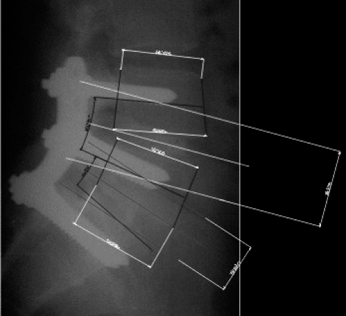 FIGURE 2.1 Digitized radiograph with DCRA landmarks and measurements given by AutoCad 2000 software (Autodesk, Inc., San Rafael, CA). |
Two independent observers, using the validated distortion-compensated method(18) measured lordosis, disc height (DAX), and dorso-ventral displacement (D) of all the unfused segments on digitized standing lateral radiographs taken immediately before surgery and after a mean follow-up of 7.51 years (range 4 to 11.5 years) (Fig. 2.1). The collected data were analyzed along with spinopelvic balance and sagittal angular parameters at follow-up.
Results
Related posts:
 Adjacent Level Disc Biomechanics
 What is the Evidence Base for IDET?
 Total Lumbar Disc Arthroplasty: Overview of Clinical Results for Existing Implants
 Functional Lumbar Artificial Nucleus Replacement—DASCOR
 A New Approach to Lumbar Disc Prosthesis
 Posterior Nonfusion Stabilization of the Degenerated Lumbar Spine with Cosmic
Stay updated, free articles. Join our Telegram channel

Full access? Get Clinical Tree


