♦ Preoperative
Operative Planning
- Physical exam
- Clinical features include palpable ridge along sagittal suture, biparietal narrowing, occipital bossing
- Closed fontanelle does not indicate sagittal synostosis (or any craniosynostosis). Craniosynostosis is a problem of the sutures, not the fontanelles.
- Much more common in boys than girls (8:1)
- Clinical features include palpable ridge along sagittal suture, biparietal narrowing, occipital bossing
- Review imaging
- Three-dimensional computed tomography scan (only in cases that the diagnosis is not clear on physical exam)
- Look for fused sagittal suture; plain films are not helpful in ruling in or out sagittal synostosis
- Three-dimensional computed tomography scan (only in cases that the diagnosis is not clear on physical exam)
- Reconstructive procedure, discuss risks/benefits with family
- Ophthalmologic consult to rule out papilledema if the family elects not to perform surgery. Elevated intracranial pressure is rare in single suture craniosynostosis but has been reported.
- Discuss timing of surgery; ideally the infant should be between 6 to 8 weeks because the earlier the surgery the better the reconstructive result. After 8 months a cranial vault reconstruction procedure may be required with the plastic surgery craniofacial team.
- Discuss donor-directed blood
Special Equipment
- Gel rolls for chest and hips
- Horseshoe head holder
- Hudson brace hand drill or perforator with pediatric burr
- Local anesthetic (bupivacaine 0.25% with 1:200,000 epinephrine, 1 mL/kg maximum dose)
- FloSeal or equivalent (thrombin with Gelfoam)
Anesthetic Issues
- Intravenous (IV) antibiotics (cefazolin 25 mg/kg/dose, clindamycin 5 to 13 mg/kg every 8 hours if penicillin allergy) given 30 minutes prior to skin incision
- Heating lamps and warming blankets to prevent hypothermia
- Arterial line
- Adequate IV access to ensure rapid administration of blood products
- Begin blood transfusion at skin incision. Typically two units of packed red blood cells are reserved: donor-directed or blood bank blood.
- Arterial line
♦ Intraoperative
Positioning
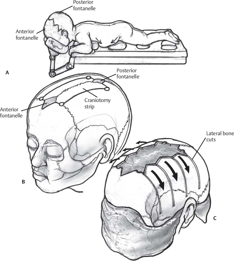
- Patient prone on horseshoe with supporting chest and hip rolls make sure there is no excess pressure on the eyeballs (Fig. 163.1A)
- Patient’s head is extended to allow easier exposure of the anterior fontanelle
- Minimal reverse Trendelenburg
- Minimal hair shave
- Hair clippers (hair is saved and given to the parents if first haircut)
Sterile Scrub and Prep
- Clean incision line and surrounding area with 70% ethanol followed by a prescrub with scrub brush followed by a two-step Betadine prep, first with Betadine soap followed by Betadine scrub
- Patient is draped with a U-shaped drape; no occlusive Ioban dressing is used
Incision and Exposure
- A biparietal sinusoidal or zigzag incision is marked midway between the anterior fontanelle and posterior fontanelle (Fig. 163.1A)
- The skin is infiltrated with local anesthetic (bupivacaine 0.25% with 1:200,000 epinephrine, 1 mL/kg is maximum dose)
- No. 15 blade knife is used to make the incision down to but not through the pericranium
- Hemostasis is obtained by insulated Bovie and bipolar cautery
- Subgaleal dissection using the Bovie on coagulation setting leaving the pericranium attached to decrease blood loss
- The exposure is complete once the anterior fontanelle and the posterior fontanelle are adequately exposed. If the patient has an excessively protuberant occiput, the dissection is performed until the occiput is exposed. Posterolaterally expose down to the asterion. Anterolaterally expose to the pterion and the squamous temporal bone.
- Wet sponges are placed over the skin flaps
- Sagittal strip craniectomy (Fig. 163.1B)
- The Bovie is used to score the areas of the initial cuts, these include burr holes placed at the asterion. If the anterior fontanelle is closed burr holes may need to be placed bilaterally at the coronal suture.
- The Hudson brace hand drill is used to make the posterior burr holes at the asterion so that the sinus is not inadvertently encountered.
- A Penfield no. 3 is used to meticulously separate the dura from bone working from the posterior burr. A Kerrison punch (2- or 3-mm) is used sometimes to enlarge the burr hole.
< div class='tao-gold-member'>Only gold members can continue reading. Log In or Register to continue
Stay updated, free articles. Join our Telegram channel
- The Bovie is used to score the areas of the initial cuts, these include burr holes placed at the asterion. If the anterior fontanelle is closed burr holes may need to be placed bilaterally at the coronal suture.

Full access? Get Clinical Tree


