Sclerotic Skull Lesions, Multiple
Miral D. Jhaveri, MD
DIFFERENTIAL DIAGNOSIS
Common
Metastases, Skull
Less Common
Fibrous Dysplasia
Paget Disease
Rare but Important
Hyperparathyroidism (“Brown Tumor”)
Osteoma
Osteopoikilosis
Melorheostosis
Osteopathia Striata
ESSENTIAL INFORMATION
Key Differential Diagnosis Issues
Osteoblastic metastasis, especially from prostate, by far the most common cause
Helpful Clues for Common Diagnoses
Metastases, Skull
Osteoblastic or treated
Most common = prostate carcinoma
Any lytic metastasis following favorable response to treatment
Other malignancies with sclerotic metastases include breast, colon, melanoma, bladder, soft tissue sarcoma
Helpful Clues for Less Common Diagnoses
Fibrous Dysplasia
20-30% polyostotic
Frontal, sphenoid, maxillary, ethmoid bones more commonly involved
Widened diploic spaces with outer table > inner table involvement
Ground-glass or sclerotic appearance
Paget Disease
Late osteosclerotic phase
Blastic lesions, often crossing sutures
“Tam-o’-shanter” skull: ↑ ↑ Diploic space, particularly inner table
“Cotton wool” skull: Focal areas of sclerosis within previous areas of osteoporosis circumscripta
Helpful Clues for Rare Diagnoses
Hyperparathyroidism (“Brown Tumor”)
Trabecular bone resorption in cranial vault
Alternating areas of lucency and sclerosis: “Salt and pepper” appearance
Brown tumors: Can become ossified during reparative process
Osteoma
In Gardner syndrome, multiple osteomas
Round dense lesions of outer table (less common in inner table)
Colonic polyposis + soft tissue tumors (especially desmoid)
Osteopoikilosis
Sclerosing bone dysplasia
Multiple radiopaque round, oval, or lanceolate spots of ↑ radiodensity
Predilection for epiphysis/metaphysis in long and short tubular bones
Skull involvement rare
Image Gallery
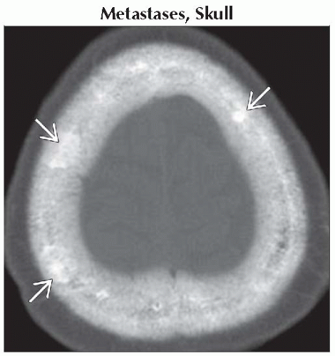 Axial bone CT shows multiple sclerotic metastases
 from prostate carcinoma. from prostate carcinoma.Related posts:Stay updated, free articles. Join our Telegram channel
Full access? Get Clinical Tree
 Get Clinical Tree app for offline access

|
