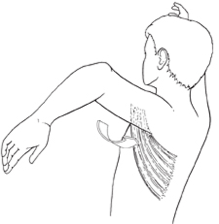
8 Secondary Reconstruction in Obstetric Brachial Plexus Injuries The patient is an 18-month-old child who suffered a left obstetric brachial plexus injury. The child was initially noted to have an internal rotation posture at the shoulder with absence of movement throughout the extremity. By 3 weeks of age, the child had regained hand grasp and extension, followed soon afterward by wrist movement in the same vectors. By the age of 3 months, the child had regained triceps extension but had 0/5 deltoid, spinatus, and biceps motor function. At the age of 6 months, the child underwent primary surgery of the left brachial plexus with excision of a nonconducting upper trunk neuroma and graft reconstruction with sural nerve. By the age of 1 year, the patient had recovered antigrav-ity biceps and deltoid strength but had also developed con-tractures in the axilla and chest referable to the latissimus dorsi, teres major, subscapularis, and pectoralis muscles. These contractures resulted in an internal rotation and adduction posture of the left upper extremity with limitation in relevant active range of motion as follows: (1) 80 degrees of shoulder abduction (normal 180 degrees), (2) 80 degrees of external rotation of the shoulder (normal 90 degrees). One of the child’s parents was an occupational therapist by training and had been supervising and performing range of motion and strengthening exercises three times a day since birth. Direct testing of the child was supplemented with viewing of videotaped therapy sessions to evaluate fully the functional limitations present during active movements of the injured extremity compared with the contra-lateral side. In addition to the restrictions in abduction and external rotation, noted too were difficulties in full active supination of the hand and the presence of the “bugler’s position” on bringing the hand to the mouth. Both signs are caused by the abnormal glenohumeral posture brought on by the described contractures. Internal rotation and adduction contracture deformity of the shoulder An internally rotated and adducted arm at the shoulder joint in a toddler with prior obstetric brachial plexus injury is the clinical hallmark. In the present case, deltoid bulk and strength were obviously present, and no electrical testing was required to confirm presence of deltoid motor function. In cases where contractures prevent abduction even to 90 degrees or where deltoid function is not clear, electromyographic (EMG) testing is appropriate to confirm the presence of functioning deltoid muscle fibers; contracture releases in the face of a completely denervated deltoid muscle are often unrewarding and must be performed with the understanding that functional improvement may not follow. Posterior subluxation and dislocation of the gleno-humeral joint due to capsule laxity often accompany the presence of internal rotation contractures. X-rays of the shoulder prior to operation will document the deformity as a baseline for future comparison, but the presence of glenohumeral instability is not a contraindication to operation. The anatomy of the glenohumeral joint is usually abnormal in children with obstetric brachial plexus injuries and allows abnormal movement vectors compared with a normal joint. Our experience with surgical management of secondary deformities related to obstetric brachial plexus injuries has evolved over the past 13 years and includes evaluation of more than 1500 patients. The most common secondary deformities of obstetric plexus injury are internal rotation and adduction of the shoulder. This probably occurs as a result of shoulder abductor and external rotator weakness due to upper trunk injury, combined with contractures of the large internal rotators and adductors (Fig. 8–1). The imbalances set up quickly and are evident within 8 to 12 months of age. Physical therapy is always the first line of management, but surgery provides a necessary tool for refractory cases. In our experience, significant imbalances still present by the age of 18 months respond more favorably to surgical management than continued therapy followed by surgery at the age of 6 to 8 years. It is unusual to see a case with late reversal of contractures and muscle imbalances by continued conservative management. This, combined with the presence of significant developmental abnormalities in the glenohumeral joint by the age of 2 years, leading to shoulder instability, indicates optimal surgical management around the age of 18 months to 2 years. In the present case, the child underwent a series of surgical procedures designed to restore shoulder muscle balance by reducing adduction and internal rotation forces and improving abduction and external rotation vectors.
Rahul K. Nath, Saleh M. Shenaq, John Laurent, Rita Lee, and Maureen Nelson
 Case Presentation
 Diagnosis
 Characteristic Clinical Presentation
 Diagnostic Tests
 Management Options
Related posts:

Stay updated, free articles. Join our Telegram channel

Full access? Get Clinical Tree


