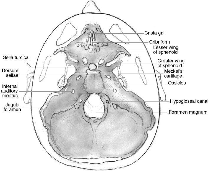
4 Skull Base Embryology • The skull base undergoes a complex sequence of developmental stages, especially in the first 12 weeks of fetal development.1–4 • The rostral neural crest cells form the whole viscerocranium and the rostral part of the neurocranium.1,2 • Migration of neural crest cells around the embryonic pharynx leads to formation of six pairs of embryonic arches. • Neural crest cells of the first two branchial arches form the bones and connective tissue of the skull. • Chondrification occurs for condensation of neural crest-derived mesenchyme. • Endochondral ossification of the skull base is predominant. • First skeletal structures to differentiate: viscerocranium, occipital bone, and cartilage of the skull base. • Skull base foramina develop as cartilage condensation around cranial nerves and blood vessels before bones are formed.2 • The chorda dorsalis is the rostral tip of the notochord, at the caudal level of the hypophysis, and represents the beginning of the skull base development. • Multiple centers of chondrification form chondroblasts around week 7: presphenoid cartilage, basisphenoid cartilage, nasal capsule, orbitosphenoid, alisphenoid, otic capsule, parachordal cartilage, occipital somites. • The ossification of the chondrobasicranium (basal plate) begins at week 8, and it is perforated by the preexisting cranial nerves and blood vessels8 (Fig. 4.1 and Table 4.1). • The parachordal cartilage forms the basioccipital elements (occipital bone and foramen magnum) at the 7th week. Exo-occipital somitic components contribute to the formation of the occipital bone and its elements, which is why the occipital bone represents a vertebral element that has expanded to support the brain.6,7,9–11 • During formation of the basioccipital elements, different developmental variants of the occipital condyles and craniovertebral ligaments may cause anatomic variants in the craniovertebral junction (e.g., “occipitalization” of the atlas, caused by ossification of the atlanto-occipital joint).12 • The ossification of the occipital bone also occurs around the hypoglossal nerves, giving rise to the formation of the hypoglossal canals. • During the formation of the skull base cartilage, the adenohypophysial pouch remains connected to the roof of the oral cavity (Rathke’s pouch). After further differentiation, the sella turcica and the sellar fossa are formed. • The presphenoid cartilages, the cartilaginous basis for the jugum of the sphenoid body, differentiate last within the medial aspect of the cranial base and bridge the gap between the postsphenoid and the cartilaginous nasal capsule (already developed by the third month of fetal development). Pearl The persistence of this communication gives rise to the craniopharyngeal canal. Table 4.1 Summary of the Primary Centers of Ossification of the Skull Base26
 Development of the Skull Base1–17:
Anatomic Part | Appearance Period of the Primary Ossification Center | Time of Fusion |
Occipital bone (basioccipital and exo-occipital parts) | 3rd embryonic month | 1–4 years of life |
Sphenoidal bone (wing, basisphenoidal and presphenoidal parts, lingula, hamulus pterygoideus) | 2nd–3rd embryonic months | From the 4th fetal month to the end of the 1st year of life |
Ethmoidal bone | 5th–6th fetal months | 6–16 years of life |
Temporal bone Pars petrosa Pars squamosa Pars tympanica |
5th–6th fetal months 3rd embryonic month 4th fetal month |
1st year of life |
Inner ear ossicles Malleus Incus Stapes |
2nd–6th embryonic months 6th fetal month 6th fetal month |
6th fetal month |
Nasal bone | 10th–11th months of life |
|
Lacrimal bone | End of the 3rd embryonic month |
|
Zygomatic bone | 3rd month of life |
|
Palatal bone | 8th–9th month of life |
|
Vomer | 3rd embryonic month |
|
• The foramen cecum represents a transient opening of the anterior skull base. At this stage, a temporary fontanel, the fonticulus frontalis, divides the inferior frontal bone from the nasal bone.13 All these structures regress during development, persisting in some pathological variants.
• The ossification of a cartilaginous bridge between the orbit and the presphenoid cartilage gives rise to the lesser wing of the sphenoid bone. Ossification of the cartilage around the optic nerve forms the optic canal.
• Around the otocyst, mesenchymal condensation leads to formation of the cochlea and semicircular canals followed by chondrogenesis around the vestibulocochlear nerve to create the internal acoustic meatus. Adjacent chondrogenesis around the carotid arteries forms the carotid canals.
Stay updated, free articles. Join our Telegram channel

Full access? Get Clinical Tree


