Spinal Cord Myelopathies

45 Spinal Cord Myelopathies


Acute Myelopathies


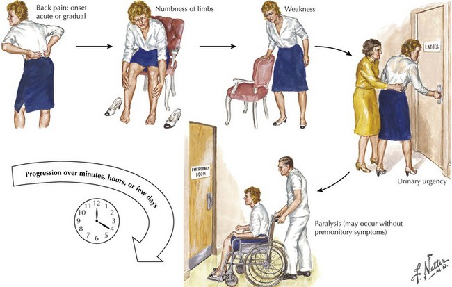
The patient presenting with an acute myelopathy provides one of the most challenging emergencies that a neurologic physician will confront. These clinical settings require expeditious and thorough evaluation including careful history, detailed neurologic examination, and immediate definitive neuroradiologic studies. In most instances, a magnetic resonance image (MRI) is the perfect study. The primary exception is the patient with a pacemaker or other medical apparatus that is not compatible with a magnet. Expeditious therapeutic intervention can sometimes reverse/stabilize a potentially disastrous clinical outcome (Fig. 45-1). Three common causes of an acute myelopathy are spinal fracture/dislocations (Chapter 60), acute transverse myelitis in young adults, and metastatic tumors in late middle- and senior-aged individuals (Chapter 53).


Acute Extradural Spinal Lesions


Trauma


Central Herniated Disc


Clinical Vignette

A 68-year-old, part-time musician suddenly fell as he was reaching for his morning newspaper while standing on an icy, hilly driveway. He was paralyzed from his neck down, and he noted numbness in all extremities. He recalled a brief involuntary ballistic movement of his right arm. A diagnosis of a brainstem stroke was made at the local hospital as it was presumed that the stroke led to his fall. Brain computed tomography (CT) was normal. Within a few days, he recovered some right-sided motor function.


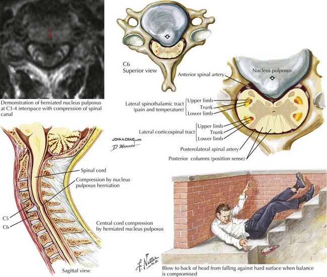
The family sought a second opinion at Lahey Clinic. Here he had a left > right quadriparesis, bilaterally brisk muscle stretch reflexes, left Babinski sign, and a midcervical right-sided sensory level for pain and temperature with preserved position sense, an exaggerated Brown–Sequard syndrome. MRI demonstrated a centrally herniated nucleus pulposus at C3–C4 with spinal cord compression, contusion, and a severe stenotic spondylotic lesion at that level. Emergency surgical decompression was performed. He had a gradual increase in function; within 1 year he could perform most activities of daily living independently, although his finger dexterity for clarinet playing was not back to his preinjury level.


In retrospect, he had sustained a syncopal event secondary to a cardiac arrhythmia. The fall per se led to the spinal column injury, the extruded central disc and cord injury. A careful neurologic examination sorted out the site of pathology and led to the diagnostic and therapeutic triumph!


Comment: Initially, this patient was thought to have a basically untreatable brainstem stroke, that is, a lesion in an entirely different part of the neuraxis. A more careful subsequent clinical examination, lying the patient on his side and examining for a specific sensory level, was the key to diagnosis, as patients with brainstem stroke rarely have total loss of sensory function in a spinal cord–level distribution (see Figs. 44-11 and 44-12).


Acute spinal cord injuries, with subsequent paraplegia or quadriplegia, are among the most dreaded sequelae to a serious bodily injury (Box 45-1). There is a tragic propensity for traumatic myelopathies to occur among vigorous and healthy young persons. Automobile and motorcycle accidents as well as sports injuries are the most common etiologies. Gunshot wounds, whether resulting from war, accident, or assault, are another source of traumatic spinal cord injury.


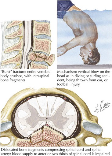
Cervical spinal fracture dislocation with resultant ligamentous tear, allowing bony fragments to directly tear or transect the spinal cord, is the common denominator in this setting (Fig. 45-2). Sometimes, there is concomitant compromise of the spinal arteries causing an associated spinal cord infarction, hematomyelia, or both. Typically, in the acute setting, spinal shock results with complete paralysis, loss of sensation, areflexia distal to the trauma site, and loss of bladder and bowel function.


Occasionally, these traumatic spinal lesions are amenable to immediate surgical correction or external traction (Chapter 60). Prognosis is always guarded. Once emergent management is completed, these patients are cared for at specialized spinal rehabilitation centers. Treatment of autonomic and sphincter dysfunction has greatly improved the long-term survival for many patients. Spinal cord repair, leading to functional recovery, is one of the greatest challenges for 21st-century neuroscientists.


Among senior citizens, unexpected falls at home, as in the above vignette, may lead to an acute central disc protrusion. Their inherent gait instability secondary to chronic neurologic or orthopedic handicaps make them susceptible to tripping on stairs, rugs, or door jams. Cardiac bradyarrhythmias leading to syncope, or epileptic seizure, with sudden loss of consciousness have a similar risk of serious spinal cord injury.


As these lesions are eminently treatable with neurosurgical intervention, consideration of these less common lesions is vital in patients who have sudden unexplained falls leading to immediate paralysis. Anatomically, the extruded disc compressed the anterior spinal cord between two vertebral bodies. Occasionally, these lesions present subacutely or chronically. Because associated bony pain or tenderness may be present, these lesions often mimic metastatic or primary tumors. However, if the patient does not have a history of malignancy, a benign mechanism, such as a central disc, must be sought (Figs. 45-3 and 45-4). Rarely, a dural arteriovenous malformation (AVM) or spinal epidural hematoma (SEH) mimics this clinical picture.


Surgery is the treatment of choice. Prognosis depends on the degree of cord compromise before surgery, the acuity of the event, the patient’s general health status, and the disc location. Cervical lesions are treated by a neurosurgeon, although a combined approach with an orthopedic surgeon is sometimes indicated. When the central disc is in the thoracic cord area, a combined neurologic and thoracic surgical approach is required.


Metastatic Malignancies


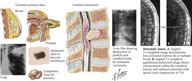
One of the most common etiologies for a nontraumatic acute or subacute extradural myelopathy occurs with various forms of metastatic carcinoma or lymphoma (Fig. 45-5). These patients initially develop spinal pain secondary to bony metastases. Within a matter of days, they begin to note symptoms of spinal cord compromise, usually with a spastic gait or bladder. Once these symptoms appear, the progression to paraplegia may be very rapid. On occasion, the spinal metastasis may be the presenting sign of a previously undiagnosed lung cancer. Emergency MRI is indicated. Treatment of metastatic extradural tumors includes radiation and sometimes surgery, chemotherapy, or both. In contrast to metastatic disease, primary intrinsic spinal cord tumors are usually either intradural extramedullary or of intramedullary origin.


Epidural Abscess


Clinical Vignette


A 60-year-old man with diabetes and a recent history of dental extractions developed lumbar and soon thereafter thoracic spine pain. During the next 2 days, his pain worsened even while lying in bed. Neither meperidine, a narcotic agent, nor a muscle relaxant relieved his pain and paraspinal muscle spasm. His difficulties rapidly worsened as he began to have trouble walking, lost sensation in his legs, and became unable to urinate.


Neurologic examination demonstrated an acutely ill febrile individual with a flaccid paraplegia, brisk muscle stretch reflexes at the knees, bilateral Babinski signs, loss of position sense in the feet and a cord level to both pin and temperature sensation at T6. There was percussion tenderness over his upper thoracic spinous processes. MRI demonstrated an extensive epidural collection extending from C6 to T10. Emergency laminectomy was performed, which demonstrated a purulent epidural abscess. Despite the emergent surgery, the patient was still significantly limited a few months later.


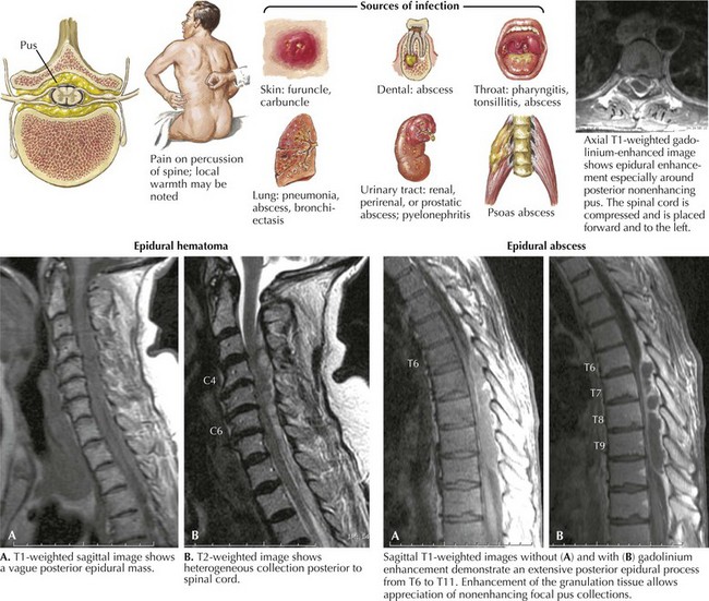
Epidural spinal abscess is a rare clinical process occurring in 2–20 cases per 100,000 hospital admissions. Its incidence appears to be increasing; these are usually seen in middle-aged adults, more often men. Despite its rarity, the potential for an epidural abscess to cause permanent paraplegia makes it one of the most urgent spinal cord emergencies (Fig. 45-6). Even though back pain is such a ubiquitous and usually benign process, it is very important for any physician to consider the potential diagnosis of an epidural abscess in every patient presenting with acute and increasing back pain, especially when febrile. The epidural space in the posterior thoracic cord is the primary site for an epidural spinal abscess to develop. These may extend to the cervical cord and rarely into the lumbar spine. Experimental studies suggest that the mass effect of the abscess, leading to cord compression, is the important clinicopathologic mechanism.


Staphylococcus aureus is the predominant etiologic microorganism leading to epidural spinal abscesses. Usually a distant septic focus provides bacterial seeding via the bloodstream, for example, skin furuncles, dental abscesses, simple pharyngitis, or a recently infected traumatic site (see Fig. 45-6). Often there is a concomitant history of diabetes mellitus, alcoholism, drug abuse, or recent spinal or extraspinal trauma. Less commonly, epidural spinal abscesses develop subsequent to vertebral osteomyelitis, pulmonary or urinary infection, sepsis, or extremely rarely bacterial endocarditis. Invasive procedures, including epidural anesthesia, spinal surgery, vascular access lines, and paravertebral injections, also provide potential mechanisms for bacterial seeding. Corticosteroid therapy may contribute to immune suppression and the possibility of secondary nosocomial infections.


Percussion tenderness over the posterior spinal processes, as well as fever, are important diagnostic clues compatible with an epidural spinal abscess Some of these individuals also develop signs of meningeal irritation such as Kernig’s sign. A rapidly developing combination of motor, sensory, and sphincter dysfunction then occurs. Often the patient becomes paraplegic and demonstrates a spinal cord sensory level. The differential diagnosis includes acute central disc, epidural metastasis often with acute pathologic fracture, and spontaneous or anticoagulation-induced hematoma.


An urgent MRI easily identifies the epidural abscess. Concomitantly, there may be a highly elevated C-reactive protein and erythrocyte sedimentation rate, often >70 mm/hour, with a modestly elevated WBC count. Emergency surgical decompression is the treatment of choice. Occasionally when no significant neurologic compromise exists, antibiotics are the primary treatment. However, careful follow-up is indicated as the patient’s clinical picture may rapidly evolve with motor and sensory loss leading to need for another MRI and surgical intervention.


Prognosis depends entirely on the patient’s expeditious presentation to a medical facility and the clinician’s level of suspicion leading to relatively early diagnosis of the epidural spinal abscess. If treatment is not initiated until after the patient becomes paraplegic, prognosis is extremely guarded.


Spinal Epidural Hematoma


Fortunately a spinal epidural hematoma is a relatively rare lesion with an acute to subacute onset. Some of these occurrences are related to anticoagulation therapy, particularly warfarin or heparin. If an anticoagulated patient sustains back or neck trauma or develops symptoms mimicking an acute meningitis that in most circumstances requires an emergent spinal puncture, and the unwary clinician has not recognized that the patient is anticoagulated, performance of a lumbar puncture (LP) is very dangerous. The patient is at risk to develop an acutely evolving paraparesis/paraplegia within 12–24 hours. By the time the effects of the anticoagulation are reversed, surgical intervention may be too late to regain neurologic function; this is truly an iatrogenic lesion that is preventable by careful assessment of all patient medications prior to performing the LP.


Spontaneous spinal epidural hematomas are being defined more frequently with the more widespread use of MRI. Very occasionally, spinal epidural hematomas seem to occur spontaneously. However, it is suggested that antiplatelet agents such as acetylsalicylic (Aspirin) or clopidogrel (Plavix) therapy may be a contributory risk factor. These lesions may present with subacutely evolving thoracic or cervical spine pain, evolving weakness, and urinary retention. Surgical decompression is the usual means of therapy, and the prognosis may be better than with those for the patient taking major anticoagulants.


Acute Intradural Intramedullary Spinal Lesions


Myelitis Secondary to Multiple Sclerosis


Clinical Vignette


Right arm weakness acutely developed in a 30-year-old woman. In retrospect, she had noted some numbness in that arm for the past 3 months, and 16 months earlier she experienced blurred vision in her right eye, with intermittent dizziness. She also had experienced episodic momentary electric shock, lightning-like sensation radiating to her buttocks often precipitated by bending her neck. Hot weather was more uncomfortable for her as she reported worsening of these symptoms and nonspecific fatigue. She had a slightly spastic left-sided gait, increased muscle stretch reflexes with a left Babinski sign, poor left-side position sense, and reduced pin and temperature sensation over her right arm and thigh.


Cervical spine MRI demonstrated an area of increased signal with ill-defined enhancement located posteriorly and on the left side of the cord (Fig. 45-7A). Brain MRI with and without gadolinium demonstrated a few periventricular lesions and other abnormalities oriented perpendicularly along vessels (Dawson fingers) within the corpus callosum. Visual evoked responses (VERs) were prolonged for her right eye. Her cerebrospinal fluid (CSF) was significant for 10 white blood cells (99% lymphocytes, 1% monocytes), and the presence of oligoclonal bands that was not identified in her serum.


The brief sensation of an electric shock running out the arms or down the back when a patient bends their neck is known as the Lhermitte sign. It is a classic symptom of cervical spinal cord posterior column pathology. Although most commonly seen in MS, because of the high incidence of this disorder, it is a non-specific symptom that is also seen in other intramedullary lesions such as vitamin B12 deficiency, or a number of extramedullary lesions causing cord compression.


Comment: This vignette presents a classic clinical picture of early multiple sclerosis (MS). The patient has an incomplete demyelinating myelitis consistent with classic hemicord syndrome, affecting ipsilateral motor and proprioceptive function and crossed sensory fibers. This is known as the Brown–Sequard syndrome. However, she also had involvement of her right arm, implying extension into the right anterior horn. The presence of the subclinical brain MRI abnormalities, as well as the prolonged VERs, point to multiplicity of demyelinating lesions in time and space consistent with the diagnosis of MS (Table 45-1).


Table 45-1 Acute Intradural Intramedullary Myelopathies


Inflammatory
Transverse myelitis


Vascular
Anterior cord syndrome

Infectious


Trauma
Central cord syndrome


Transverse Myelitis


Clinical Vignette


A 16-year-old boy suddenly began to walk with both knees in a flexed posture. Initially, his parents thought he was just joking. However, later that evening he began to experience knife-like pain in midback radiating around his ribs toward his epigastrium. The next morning, he awakened unable to get out of bed. He was unable to void. On neurologic examination he was paraplegic, his muscle stretch reflexes were absent, and his plantar response “ambiguous.” Sensory exam suggested a T10 level for both pain and temperature modalities.


Pertinent laboratory findings included CSF findings with a protein 175 mg/dL, and 30 WBC with 90% lymphocytes. Nerve conductions demonstrated prolonged F waves but otherwise normal motor and sensory nerve conductions. The spinal cord had a focal demyelinating lesion with gadolinium enhancement involving most of its transverse diameter at T9–T11. Unfortunately a course of intravenous (IV) methylprednisolone was ineffective; he remained paraplegic, with a persistent dense sensory level and ongoing incontinence.


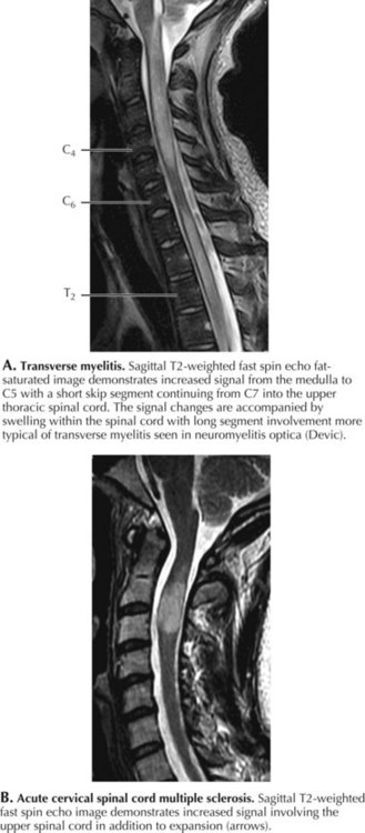
Very often, there is no underlying pathology identified with many transverse myelitis (TM) cases. Acute transverse myelitis most likely represents another type of autoimmune CNS disorder. A nonspecific “viral infection” may be the inciting mechanism or sometimes a bacterial infection, and rarely this may occur subsequent to immunization. Sometimes, a transverse myelitis is preceded by or associated with optic neuritis. This is known as neuromyelitis optica (NMO), an unusual autoimmune demyelinating disorder associated with a very specific serum autoantibody referred to as NMO-IgG (Fig. 45-7). This antibody binds to the CNS-dominant water channel, aquaporin 4; this is a structure that is normally present on astrocytes. The diagnostic criteria for neuromyelitis optica include optic neuritis, acute myelitis, and two of the following three: brain MRI not characteristic for multiple sclerosis, contiguous spinal cord MRI lesion extending over three or more vertebral segments, and NMO-IgG positive status.


Motor, sensory, and sphincter disturbances vary in degree if the process begins subacutely. However, with an acute onset, a lesion can rapidly develop, mimicking a traumatic lesion with paraplegia or quadriplegia, total sensory loss, and absence of bladder and rectal sphincter function as per the above vignette. A complete transverse myelitis interrupts all ascending tracts below the lesion, leading to a “sensory level” and concomitantly a flaccid paraplegia or tetraplegia depending on the lesion level as all descending tracts above the pathologic site, particularly the corticospinal tracts, are compromised. Over time, spasticity develops. Interruption of the anterior and lateral spinothalamic tracts and dorsal columns leads to the cord level. Pinprick or temperature sensation is the most easily localized, with loss 1–2 levels below the lesion site. Bladder and bowel functions are also impaired.


Differential diagnosis includes cord infarct, arteriovenous malformation, radiation myelopathy, metastatic or intrinsic tumors, and central cord compression for a spondylotic central disc. Very rarely, TM is part of an acute disseminated encephalomyelitis. Systemic lupus erythematosus (SLE), other types of vasculitis, sarcoidosis, Sjögren syndrome, antiphospholipid antibody syndrome, and Schistosoma mansoni parasitic infection are other predisposing mechanisms.


MRI with gadolinium is the procedure of choice to exclude compressive lesions, especially when history and exam suggest a specific level of spinal cord dysfunction (see Fig. 45-7). Brain lesions consistent with MS are demonstrated with MRI in approximately half of all TM patients. Transverse myelitis appears with T2 signal hyperintensity on MRI. The area of signal abnormality may be focal or extensive in cross section and length. Gadolinium enhancement is frequent. Cord swelling is present to variable degrees. Large cross-sectional area, multisegment length, cord expansion, and peripheral enhancement are most consistent with a diagnosis of TM. In contrast, MS lesions tend to be smaller, usually involving only 1–2 segments, and are often multifocal. Total cross-sectional area and multisegment length are uncommon in MS, although cord expansion and enhancement are frequent with larger acute inflammatory MS lesions. Gadolinium-enhanced brain MRI, and optical coherence tomography (OCT), even more than visual evoked potentials (Chapter 46), help determine the presence of multifocal demyelinating disease. Associated cerebral white matter lesions and oligoclonal bands in CSF increase the later probability of developing unequivocal MS. Oligoclonal bands, however, are nonspecific for multiple sclerosis and not always present in individuals with multiple sclerosis.


When evaluating a patient with TM for the presence of infection or systemic inflammatory disease, a lumbar puncture is indicated for CSF analysis (cell count, protein, oligoclonal bands, culture, glucose, and viral polymerase chain reactions [PCR]) and or viral titers, and serologies. Blood work required includes ESR, C-reactive protein, antinuclear antibodies, anti-DNA antibodies, SSA, SSB, anticardiolipin antibodies, lupus anticoagulant, complement, and angiotensin-converting enzyme. Viral and bacterial screen might include varicella zoster, enterovirus, Coxsackie virus, Epstein-Barr virus, cytomegalovirus, herpes simplex, hepatitis, HIV, and Lyme titers.


Methylprednisolone, 1 g intravenously for 5–10 days, is indicated in all TM patients as occasional NMO patients respond to this therapy and/or plasma exchange. However, there is always a variable degree of response. The degree of recovery depends on the rapidity of development and severity of deficit. Unfortunately, most NMO patients may have significant optic nerve and spinal cord deficits and a tendency to have severe relapses. Intermittent or chronic immunosuppressive treatment is often indicated. Such a temporal profile also occurs with SLE, antiphospholipid antibody syndrome, and vascular malformations of the cord. In contrast, a relapsing and recurring transverse myelitis is rare when TM is not a manifestation of either NMO or MS.


Spinal Cord Infarction/Ischemic Myelopathy


Anterior Spinal Artery Syndrome


Clinical Vignette

A vigorous 56-year-old state police officer was found to have an extensive thoracoabdominal aneurysm requiring heroic surgical repair with replacement of his entire descending aorta from its arch in the chest to the distal bifurcation within the abdomen. Although the primary surgical procedure appeared to be successful, when the patient awakened he was unable to move his legs or empty his bladder and he had numbness distal to T8–T10. Neurologic examination demonstrated the patient to be paraplegic but with preserved strength in his arms and upper body, loss of temperature and pain sensation, with a vague numbness from his upper abdomen to the tips of his toes, and preserved position and vibratory sensation.


Thoracic and cervical MRI performed within a few hours of his awakening was normal. This excluded a mass lesion potentially capable of being surgically treated but could not reveal an early spinal cord infarct. The patient’s clinical course was otherwise stable. When there was no improvement in his neurologic status, he was transferred to a spinal rehabilitation unit.


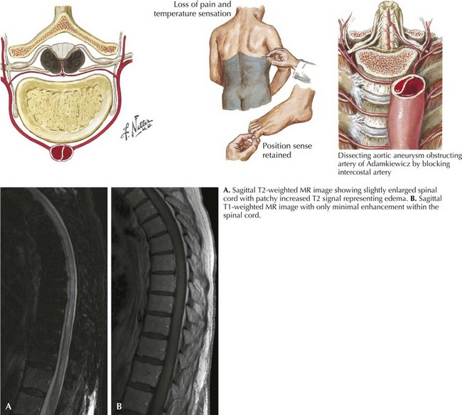
The clinical picture of paralysis secondary to loss of corticospinal tract function and sensory change from impaired spinothalamic function, as well as infarction of the anterior horn cells, but with very preserved posterior column function, is classic for an anterior spinal artery distribution spinal cord infarction (Fig. 45-8). Onset of anterior spinal artery syndrome, although usually sudden, may occasionally be gradual over hours or days.


Because of the significant collateral circulation, spinal cord infarction is very much less common than cerebral ischemia; thus spinal cord infarction and transient ischemic attacks involving the spinal cord rarely occur. This diagnosis must be considered in patients who present with sudden onset of nontraumatic weakness, a sensory loss with a definable level to pain and temperature with preserved posterior column function, and bladder dysfunction. Paresthesia or radicular pain can occur at the infarct level. Patients may develop either a bilateral flaccid paraplegia or quadriplegia depending on the site of cord occlusion. At the cervical level, the arms are flaccid and eventually atrophic because of anterior horn involvement, although the legs become spastic. Thoracic cord infarcts lead to a spastic paraplegia. Initial areflexia changes to hyperreflexia with the presence of Babinski signs.


Spinal cord infarction is typically secondary to inadequate arterial flow through the anterior spinal artery, which supplies the anterior funiculi, anterior horns, base of the dorsal horns, and anteromedial aspects of the lateral funiculi. Thus, the corticospinal and spinothalamic tracts are affected bilaterally. The upper to midthoracic spinal cord is poorly vascularized, and these watershed zones are more susceptible to infarction. Interruption of blood supply from the aorta to the intramedullary spinal vasculature can cause infarction. In contrast, a posterior spinal artery infarction is rare because of well-developed collaterals.


Spontaneous dissecting aneurysms of the descending thoracic or upper abdominal aorta can occlude the ostia of segmental spinal arteries; atherosclerosis of the aorta and its branches, and iatrogenic ischemia from recent aortic surgery are the usual pathophysiologic mechanisms predisposing to spinal cord infarction. Aortic surgical procedures may reduce vascular supply to the radicular and spinal arteries. Procedures such as thoracotomy and nephrectomy sometimes compromise intercostal or lumbar artery flow, which give rise to radicular arteries. The presence of concomitant chest or abdominal pain, limb ischemia, or loss of peripheral pulses suggests a possible aortic dissection.


Very rarely emboli or arteritis may be responsible for an intramedullary cord infarction. Emboli to the anterior spinal artery may be derived from atheromatous, septic, fibrocartilaginous, and air (decompression illness/caisson disease) sources. Vascular angiitis with subsequent thrombosis from primary CNS vasculitis, syphilis, tuberculosis, sarcoidosis, and schistosomiasis can all cause cord infarction.


Chronic Myelopathies


Extradural Myelopathies


Cervical Spondylosis


Clinical Vignette


An obese septuagenarian, with previously diagnosed diabetic polyneuropathy manifested by burning discomfort in his feet, presented with a 4- to 6-month history of increasing leg numbness. These new symptoms were totally different from the mild tingling and burning that had been chronically present for the past 10 years. He began to require a cane to maintain his equilibrium when walking. Although he initially tolerated the newer symptoms, he began to be concerned that he could not walk safely without relying on a walker. He sought further medical opinion. He previously had a myocardial infarction.


Neurologic examination demonstrated a broad-based, spastic gait, brisk muscle stretch reflexes, and bilateral Babinski signs. Pinprick and temperature sensation were reduced in a stocking-glove distribution in his legs. There was a question of a bilateral cord level to pin sensation at C-7. Position sense was absent at the toes, and vibratory sense lost at the ankles.


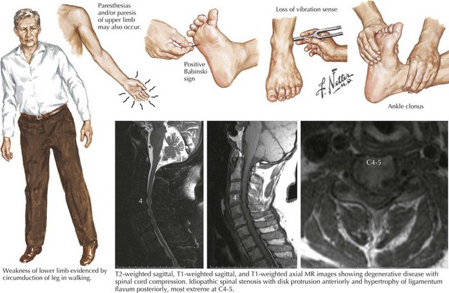
MRI revealed spinal stenosis and cord edema at C5–C6. He had severe spinal stenosis with multilevel spondylosis, disc protrusion, and end-plate osteophytes. After a 3-month period of observation, his gait difficulties increased. A cervical posterior laminectomy was performed. Subsequently, after a period of rehabilitation hospitalization, he gradually regained the ability to walk independently.


One needs to always carefully evaluate the patient with a chronic primary sensory polyneuropathy who begins to develop disproportionately increased gait difficulty. Cervical spinal stenosis is a common chronic disorder. As occurred in this instance, one may define a quite remediable condition.


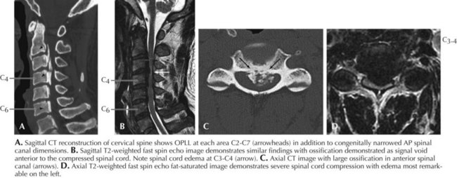
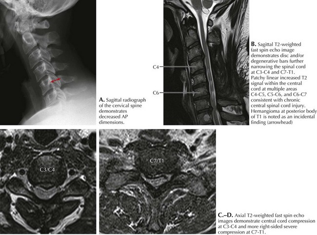
Spondylosis, a normal aging process, is the most common cause of a cervical myelopathy (Figs. 45-9 and 45-10; Box 45-2). This results from disc degeneration followed by reactive osteophyte formation, fibrocartilaginous bars, spondylotic transverse bars, articular facet hypertrophy, and thickening of the ligamentum flavum causing spinal canal narrowing. Subsequently, gradual spinal cord compression may occur; it is particularly likely in patients having congenitally narrowed spinal canals. In its simplest form, a chronically herniated central nucleus pulposus in patients with congenital stenosis can produce a cervical myelopathy. Although many senior individuals have radiographic signs of cervical spondylosis, most are asymptomatic. When a myelopathy develops, clinical findings can present acutely, subacutely, or over many years. Sometimes both a cervical myelopathy and adjacent radiculopathy may occur in the same spondylotic patient.

Pathophysiology and Etiology


Typically, the spinal canal is 17–18 mm in diameter between C3 and C7. A narrower cervical spinal canal may range from 9 to 15 mm; however, a compressive spondylitic myelopathy rarely occurs when the canal diameter is >13 mm. Cervical cord diameter ranges from 8.5 to 11.5 mm, averaging approximately 10 mm. Disc protrusion and other reactive and degenerative processes further reduce canal dimension. Direct cord compression, compromised blood supply to the cord or venous stasis, and other mechanical factors, such as rheumatoid arthritis, can in combination or independently cause irreversible damage.


Normally, the spinal cord moves cephalad and posteriorly within the canal during neck flexion and caudally and anteriorly during neck extension. If osteophytes, discs, and hypertrophied ligaments make contact with the cord, the cord sustains additive trauma, leading to development of a clinical myelopathy. The disc levels affected are C5–C6, C6–C7, and C3–C4, in order of their clinical frequency.


In this setting, the spinal cord may become pathologically, grossly flattened, distorted, or indented. Demyelination of the lateral columns occurs at the lesion site with consequent lateral column degeneration below the lesion. Concomitant dorsal column degeneration occurs at and above the damaged segment(s). There may also be damage and loss of nerve cells in gray matter. Ischemic changes, gliosis, demyelination, and even cavitation necrosis sometimes also result.


Jun 4, 2016 | Posted by in NEUROLOGY | Comments Off on Spinal Cord Myelopathies

Full access? Get Clinical Tree

Get Clinical Tree app for offline access