♦ Preoperative
Operative Planning
- Review imaging: magnetic resonance imaging (MRI), myelography (optional), spinal angiography
Embolization
- Embolization as sole intervention associated with high rate of recurrence
- In patients with acute neurologic decline without hemorrhage (Foix-Alajouanine syndrome), embolic occlusion of fistula at conclusion of diagnostic selective angiogram prior to surgery may prevent further deterioration
- Give dexamethasone postembolization to reduce swelling
Routine Equipment
- Laminectomy instruments
- High-speed drill (optional)
- Microsurgical instruments
Special Equipment
- Consider neurophysiological monitoring of somatosensory evoked potentials (SSEPs) and motor evoked potentials (MEPs)
Operating Room Set-up
- Open-frame spinal table or electric table with bolsters or Wilson frame
- Ensure ability to obtain anteroposterior and lateral radiographs to confirm operative levels
- Headlight
- Loupes (optional)
- Bipolar and Bovie cautery
- Microscope with bridge
- General anesthesia
- Arterial line for blood pressure monitoring
- Intravenous antibiotics (cefazolin 2 g or vancomycin 1 g for adults) should be given 30 minutes prior to incision
- Minimize halogenated inhalational agents and nitrous oxide if monitoring SSEPs and MEPs
♦ Intraoperative
Positioning
- Patient prone
- Open-frame spinal table or electric table with bolsters or Wilson frame
- Ensure ability to obtain anteroposterior and lateral radiographs to confirm operative levels
- For lesions at T6 or above, pad arms well and tuck along sides; for more distal lesions, abduct shoulders and flex elbows 90 degrees
- Secure head with foam mask, Gardner-Wells tongs with 15 lb of inline traction, or Mayfield head holder
- If using foam mask, ensure no ocular pressure
Sterile Scrub and Prep
- As for posterior cervical or posterior thoracic approach
Incision
- Center midline linear incision over levels of the lesion to permit exposure one level proximal and one level distal to the lesion
Laminectomy
- Bilateral subperiosteal exposure to medial facet joints bilaterally
- Perform bilateral laminectomies from one level proximal to one level distal to the dural arteriovenous fistula (AVF)
- Do not violate facet joints (may lead to postoperative kyphotic deformity)
- Wax bone edges, obtain meticulous epidural hemostasis
Dural Opening
- Open dura in midline
- Secure edges of dura to paraspinal muscles with 4–0 silk tacking sutures
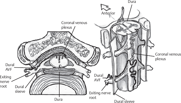
Interruption of Dural Arteriovenous Fistula (Fig. 138.1)
- Retract dura laterally to allow dissection of arachnoid around the underlying vessels
- Carefully identify medullary draining vein by inspection and comparison with angiogram
< div class='tao-gold-member'> Only gold members can continue reading. Log In or Register to continue


