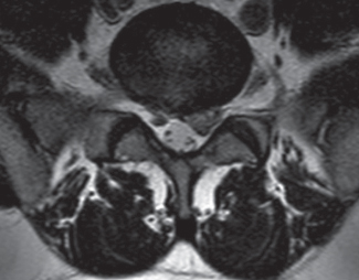
45 What are the ligamentous structures that connect C1 to the occipital bone and where do they attach?1 1. Anterior atlanto-occipital membrane from the anterior margin of the foramen magnum to the anterior arch of C1 2. Posterior atlanto-occipital membrane from the posterior margin of the foramen magnum to the posterior arch of C1 3. The superior attachment of the cruciate ligament What are the ligamentous attachments of C2 to the occipital bone? 1. The tectorial membrane running from the posterior aspect of the body of C3, C2, and the dorsal aspect of the odontoid process to the anterior aspect of the foramen magnum 2. The alar ligament from the lateral aspect of the odontoid process to the occipital condyle on each side 3. The apical ligament between the superior aspect of the odontoid process and the foramen magnum What are the ligaments connecting C1 and C2? 1. The transverse ligament (which also represents the horizontal part of the cruciate ligament) from one lateral mass of the atlas to the other embracing the posterior aspect of the odontoid process 2. The descending portion of the cruciate running behind C2 vertebral body At what cervical level can the carotid artery be compressed and where?2 • At the level of the sixth cervical vertebra, at the level of the anterior tubercle of the transverse process (Chassaignac tubercle or tuberculum caroticum). • This tubercle separates the vertebral artery from the carotid. • It is also a very important landmark for regional anesthetic blocks. What is the lateral recess?3 It is a triangular space formed by the posterior aspect of the vertebral body, the pedicle, and the superior articular facet. How can the lateral recess contribute to radicular symptoms?4 In a different situation where the lateral recess size is less than 3 mm, including facet hypertrophy with or without a herniated disk and spinal canal stenosis, the nerve exiting at this spinal level can be compressed. Fig. 45.1 Axial T2-weighted MRI of the lumbar spine demonstrating lateral recess stenosis secondary to a herniated disk. Define the vertebral lamina, • Laminae are two plates of bone extending from the pedicle in a posterior-medial direction to close the vertebral arch. • They provide attachment to the yellow ligament (ligamentum flavum). • Their width and orientation in the sagittal plane vary throughout the spine. What forms the neural foramen in the cervical spine? • The neural foramen is formed: • Superiorly and inferiorly by the pedicle of the vertebras above and below • Posteriorly by the facet joint • Anteriorly by the vertebral body and disk. What is the direction of the pedicles throughout the lumbar spine?5
Structural and Functional Anatomy
45.1 Structural Anatomy
Related posts:


Stay updated, free articles. Join our Telegram channel

Full access? Get Clinical Tree


