Subarachnoid Hemorrhage

57 Subarachnoid Hemorrhage


Subarachnoid hemorrhage (SAH) refers to bleeding beneath the arachnoid coverings of the brain surface and within the contained cisterns. The incidence is about 6–8 per 100,000 in most countries in the northern hemisphere and is highest between the fifth and seventh decades of life. The multiple etiologies for SAH are classified into primary aneurysmal and spontaneous nonaneurysmal mechanisms. Ruptured intracranial aneurysm is the most preponderant cause of spontaneous SAH, accounting for up to two thirds of all cases. One study of acute SAH in more than 6300 patients demonstrated that 51% of the patients had ruptured intracranial aneurysm. Nonaneurysmal SAHs include arteriovenous malformations (AVMs), angiographically occult vascular malformations (cavernous malformation or angioma), idiopathic and iatrogenic coagulopathies, bacterial endocarditis, venous thrombosis, inflammatory processes such as granulomatous angiitis, arterial dissections, occasional tumors, hypertension, and drug abuse. In addition, pathologic processes within the spinal canal, such as spinal AVMs and spinal neoplasms, especially myxopapillary ependymomas, can rarely lead to SAH. Despite the normal neurologic examination results and head CT in the vignette, the patient’s history was so compelling for a ruptured aneurysm or warning leak that her neurologist proceeded with the evaluation to find a large treatable berry aneurysm.


Subarachnoid hemorrhage is a catastrophic neurologic event having a precipitous onset, frequently without any premonitory warning. In North America, 28,000 patients per year experience a ruptured aneurysm. Slightly more than half die shortly after rupture. Among those who survive to reach a hospital, there is an additional 20–25% chance of further ruptures within the first 2 weeks, and the overall mortality during the first month is approximately 50%. Of those patients who survive, only about 30% will have a favorable outcome.


Aneurysmal SAH, although catastrophic, can often be treated successfully. When an aneurysm is identified before rupture, treatment can be curative, preventing the devastating effects of a SAH. Recognition of SAH, accurate diagnosis, and timely treatment are essential.


Crucial points in the history of patients with a recent headache are the abruptness of pain onset and the severity of discomfort. Lack of abnormality on the neurologic examination does not exclude a symptomatic aneurysm, and therefore, a detailed history and careful evaluation of such patients is essential. Furthermore, a mild hemorrhage as in the first vignette, may not be observed on CT after just 24 hours. Therefore, in spite of a negative head CT, cases in which there is still a high suspicion of a ruptured aneurysm would require angiography to identify the aneurysm and avoid rebleeding with its associated 50% mortality rate.


Clinical Presentation


The classic symptom of SAH is the “worst headache of one’s life.” Headaches associated with aneurysm rupture are frequently sudden in onset and often described as a severe thunderclap, excruciating and unbearable. The headache peaks rapidly and is frequently associated with pain extending across the head and toward the neck. The headache is usually global, with a constant viselike ache but occasionally throbbing. Unilateral aches or a retro-orbital stab-like pain, even fleetingly, raise the suspicion of a possible posterior communicating artery aneurysm.


Nausea and vomiting, neck pain, and altered consciousness are often associated with the headache. Approximately 30% of patients are found to be confused and lethargic after the ictus. During the moment of rupture, one fourth of patients become comatose and up to 40% have transient loss of consciousness.


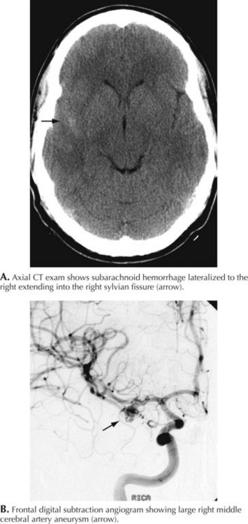
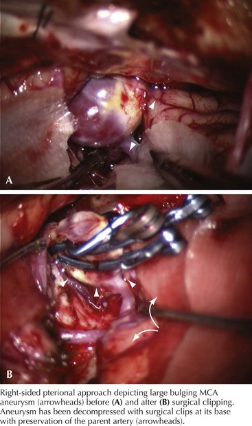
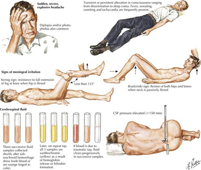
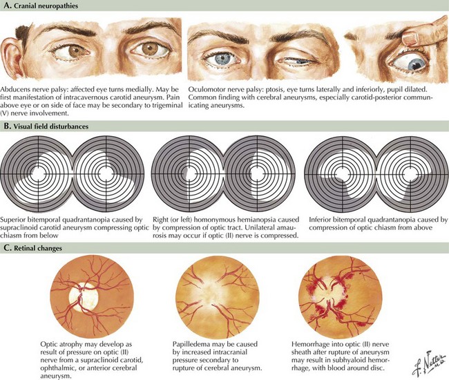
Seizure-like activity may be observed. The incidence of true seizure activity in patients with SAH is estimated at 20%. Seizures in SAH are most commonly associated with middle cerebral artery (MCA) and anterior communicating artery (ACA) aneurysmal rupture causing intracerebral hematomas. Unfortunately, many patients recall having a sentinel hemorrhage or warning leak with a fleeting but severe headache within the 2–3 weeks before the major ictus. This headache is somewhat milder and usually not associated with meningismus; it is often ignored until the catastrophic return of a major rupture. When evaluating patients with SAH or sudden severe headache, special attention should be focused on the level of consciousness, focal neurologic signs such as hemiparesis or cranial nerve palsies, and signs of meningismus (Fig. 57-5). Meningismus frequently occurs, associated with nuchal rigidity. Brudzinski’s maneuver is an excellent means of evaluating meningismus; the examiner flexes the patient’s neck, precipitating hip flexion, knee flexion, and hamstring pain. Diplopia (due to abducens or oculomotor nerve palsies) and visual loss (chiasmal or optic nerve involvement) are caused by either cranial nerve compression from the aneurysmal dome or aneurysmal rupture and increased intracranial pressure (Fig. 57-6).


Examination of the optic fundi frequently discloses retinal or preretinal hemorrhage, subhyaloid hemorrhages, and occasional papilledema. Hemorrhage into the vitreous results in Terson syndrome, with scarring and epiretinal membrane formation (macular pucker) and eventually visual loss or distortion. Terson syndrome is a frequent cause of visual loss in SAH, which often goes unnoticed until the patient regains consciousness 1–2 weeks later. It is often related with more severe subarachnoid bleeds that cause loss of consciousness and papilledema. Its association with ACA aneurysms is less clear. The long-term prognosis for vision in this situation is fairly good; however, a vitrectomy is occasionally required.


When the aneurysm ruptures and dissects into adjacent brain tissue, various focal deficits may also be found on examination.


Differential Diagnosis


Patients presenting with a sudden apoplectic-type headache with associated meningismus or altered mental status must be considered to have an SAH until proven otherwise. However, SAH symptoms are sometimes confused with other disorders, including migraine headaches, hypertension, meningitis, cervical spine disorders, vertigo, and syncope. The various vascular headache syndromes remain the most common mimics.


Although migraines are often characterized by the patient as sudden, a careful history reveals that they typically have a gradual onset, with progression over minutes to hours, at times, to the degree of excruciating pain often with nausea and vomiting. Many are preceded by a classic visual aura of fortification spectra or scintillating lights gradually evolving then regressing over minutes before the headache occurs.


Cluster headache is another benign but severe headache syndrome with a well-defined clinical presentation. These headaches typically affect men, awakening them from sleep with a terrible unilateral periorbital and frontal pain. Cluster headaches are almost always associated with unilateral conjunctival injection, excessive lacrimation, and nasal stuffiness. They have a limited time course, usually lasting 45–60 minutes. They occur nightly in a temporal cluster for 6–8 weeks but may recur several times within a day. When this pattern is established, the diagnosis is secure. However, when the patient first experiences this headache in early midlife, a careful evaluation is indicated to exclude SAH. A therapeutic response to inhalation of 100% oxygen is diagnostic.


Paroxysmal hemicrania is a related disorder with an equal sexual distribution. Its response to indomethacin is a specific therapeutic diagnostic modality.


Orgasmic postcoital or exercise-induced headaches are another group of benign headaches that occur during sexual intercourse or with significant exercise. Those related to sexual activity generally occur precipitously at the peak of orgasm. These incapacitating severe headaches mimic the onset of an acute SAH and require the same full evaluation to exclude a ruptured aneurysm as other patients presenting with spontaneous sudden first-time severe headaches. Orgasmic, postcoital, or exercise-induced headaches are essentially diagnoses of exclusion.


Diagnostic Approach


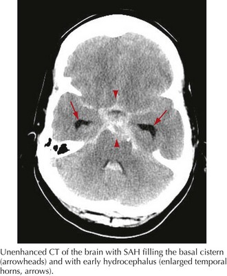
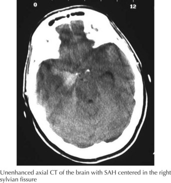
The clinical diagnosis of SAH is best confirmed with brain CT (Fig. 57-7). Its sensitivity is highest in the first 24 hours after headache onset. A mild hemorrhage may wash away within 24 hours but approximately 50% of severe SAHs are still visible on CT 1 week after the ictus, and only one third are seen after 2 weeks. CT confirms the presence of SAH and frequently highlights associated issues such as hydrocephalus, intraparenchymal hematoma, intraventricular hemorrhage, or subdural hemorrhage.


Whenever the clinical suspicion of SAH exists but CT is negative, a lumbar puncture must be performed. A nontraumatic tap is crucial. When the presence of blood in the CSF does not clear between the first and fourth tubes, this is particularly suggestive of SAH (See Fig. 57-5). However, a more sensitive indicator is CSF xanthochromia, which represents lysis of erythrocytes with degradation of heme products into bilirubin within the CSF. This frequently renders the CSF a yellowish color within 1–3 hours after an SAH, and often persists for approximately 2–3 weeks.


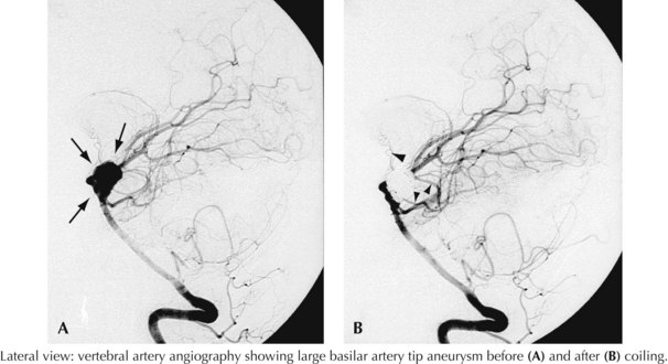
When SAH is confirmed by CT or lumbar puncture, the cause of the hemorrhage is best evaluated with a four-vessel cerebral arteriogram. An aneurysmal source is found in 80–85% of arteriograms preformed for suspected SAH. If arteriography is negative after SAH, a repeat study should be performed approximately 10 days later. Although reliance on CT angiography rather than catheter angiography has been increasing, cerebral arteriography remains the accepted standard for evaluating patients with SAH.


To ensure proper communication, predict outcomes, and guide management, a clinical grade for each SAH is needed. Several grading scales are available; the most widely used is the Hunt–Hess scale—a five-tiered description of the patient’s state and an indicator of prognosis (Table 57-1).


Table 57-1 Hunt–Hess Grading Scale for Berry Aneurysms


Only gold members can continue reading. Log In or Register to continue

Stay updated, free articles. Join our Telegram channel

Jun 4, 2016 | Posted by in NEUROLOGY | Comments Off on Subarachnoid Hemorrhage

Full access? Get Clinical Tree

Get Clinical Tree app for offline access
Grade Description
1 Asymptomatic, or mild headache and slight nuchal rigidity
2 Moderate to severe headache, nuchal rigidity, no neurologic deficits other than cranial nerve palsies.
3 Mild focal deficit, lethargy, confusion
4 Stupor, hemiparesis, central neurologic signs
5