The eyes and visual system
An examination of the eyes and the visual system can prove helpful in patients with no visual or ocular symptoms. This section will first describe examination of the eye generally, then examination of the pupils, visual function, acuity, visual fields and fundoscopy. Eye movements are discussed on pages 18–19.
General examination of the eye
Ptosis is common and is often missed (Table 1). Partial ptosis is usually associated with unilateral overactivity of the frontalis. Ptosis is not a feature of facial nerve palsy (with a facial nerve palsy the eye does not close).
| Ptosis | Causes | Features |
|---|---|---|
| Neurogenic | ||
| Neuromuscular junction | Myasthenia gravis | Variable ptosis that fatigues, may be associated diplopia and facial weakness |
| Myopathic | Myopathies, especially myotonic dystrophy | Usually symmetrical. Features of associated muscle disease |
| Mechanical | Aponeurotic dehiscence (common in the elderly) | The tarsal plate is separated from the levator muscle |
Remember false eyes can be cosmetically effective – a pitfall in exams.
Examination of the pupils
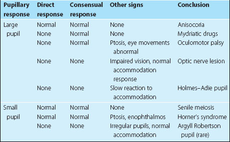
Ask the patient to look into the distance and shine a light twice in each eye in turn. First, look at the response in the eye into which you are shining the torch (the direct response), and then at the response in the other eye (the consensual response). Then ask the patient to look at your finger held 15 cm from the patient’s face and look at the pupils for brisk constriction – the accommodation response. If no response is obtained from shining a light into the eye, but there is a normal response on accommodation, this is called an afferent pupillary defect and indicates significant optic nerve disease. Other abnormalities are summarized in Table 2.
Stay updated, free articles. Join our Telegram channel

Full access? Get Clinical Tree


