Fig. 15.1
Thorotrastoma. The patient is an 82-year-old female with right vocal cord paralysis and a history of Thorotrast extravasation during an angiogram performed in the 1950s. Axial (a, b), sagittal (c), and coronal (d) CT images demonstrate hyperattenuating material surrounding the right carotid sheath, extending superiorly into the retropharyngeal space to the level of C1 and extending inferiorly to the mediastinum
15.4 Differential Diagnosis
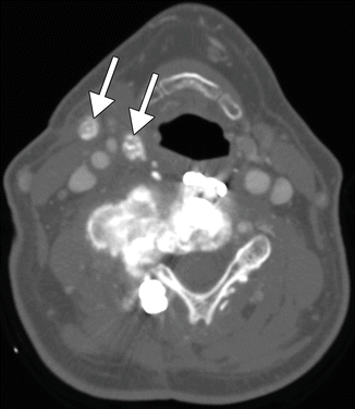
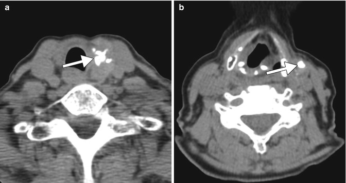
The findings in cervical Thorotrast extravasation are rather characteristic and unusual. Conditions that can potentially resemble Thorotrast deposits in the neck on CT are heterotopic ossification, osteosarcoma metastases (Fig. 15.2), treated lymphoma with calcified lymph nodes, and other tumors that have a propensity to calcify, such as papillary thyroid carcinoma (Fig. 15.3), and metal clips (Fig. 15.4).


Fig. 15.2
Osteosarcoma metastases. Axial CT image shows an osteoblastic mass arising from a cervical vertebra in a previously operated site. There are also hyperattenuating right cervical lymph node metastases (arrows)

Fig. 15.3


Papillary thyroid carcinoma. Axial CT image (a) shows a calcified mass in the left thyroid lobe (arrow). Axial CT image at another level (b) shows a calcified nodal metastasis (arrow)
Stay updated, free articles. Join our Telegram channel

Full access? Get Clinical Tree


