♦ Preoperative
Imaging
- Magnetic resonance imaging (MRI) to assess spinal cord compression and extent of pathology (soft tissue and pleural extension)
- Plain x-rays to evaluate alignment, count ribs, and identify natural fiducials which may facilitate correlation of intraoperative imaging findings with MRI images
Preoperative Care
- Somatosensory and motor evoked potentials as indicated
- Pulmonary function tests if single lung ventilation may be required (typically above T8)
Equipment
- Self retaining rib retractor
- Extended length Bovie may be useful
- Long handled Kerrisons, pituitaries, and curettes
- Long Frazier suction tips may be useful
Operating Room Set-up
- Double lumen endotracheal tube or bronchial obturator may facilitate single lung ventilation
- Somatosensory and motor evoked potential monitoring (optional)
- Right lateral decubitus position allows for left-sided thoracotomy, which is preferred because of relative ease of mobilizing the aorta versus vena cava
- Right (typically) lateral decubitus position on operating table (bottom leg bent, top leg straight, pillow between the knees)
- Consider use of beanbag to secure patient in lateral position with care to pad pressure points, especially dependent areola
- Drape iliac crest into field in case autograft is needed
- Safety straps at shoulder, thigh, and calf levels to allow for safe table rotation as needed during the exposure, decompression, and reconstruction
- Axillary roll
Exceptions to Left Thoracotomy Preference
- Patients who cannot tolerate left lung deflation (but can tolerate right lung deflation)
- Pathology eccentric to the right (dumbbell neurofibroma, vertebral tumor extending into the right chest, and rightward eccentric intraspinal canal pathology)
- Patients with flank skin lesions precluding left-sided approaches (bruising, psoriasis, etc.)
- History of prior transthoracic spinal surgery from the right (it is preferable to deal with pleural scarring rather than to risk spinal cord infarction taking both segmental vessels at the same level)
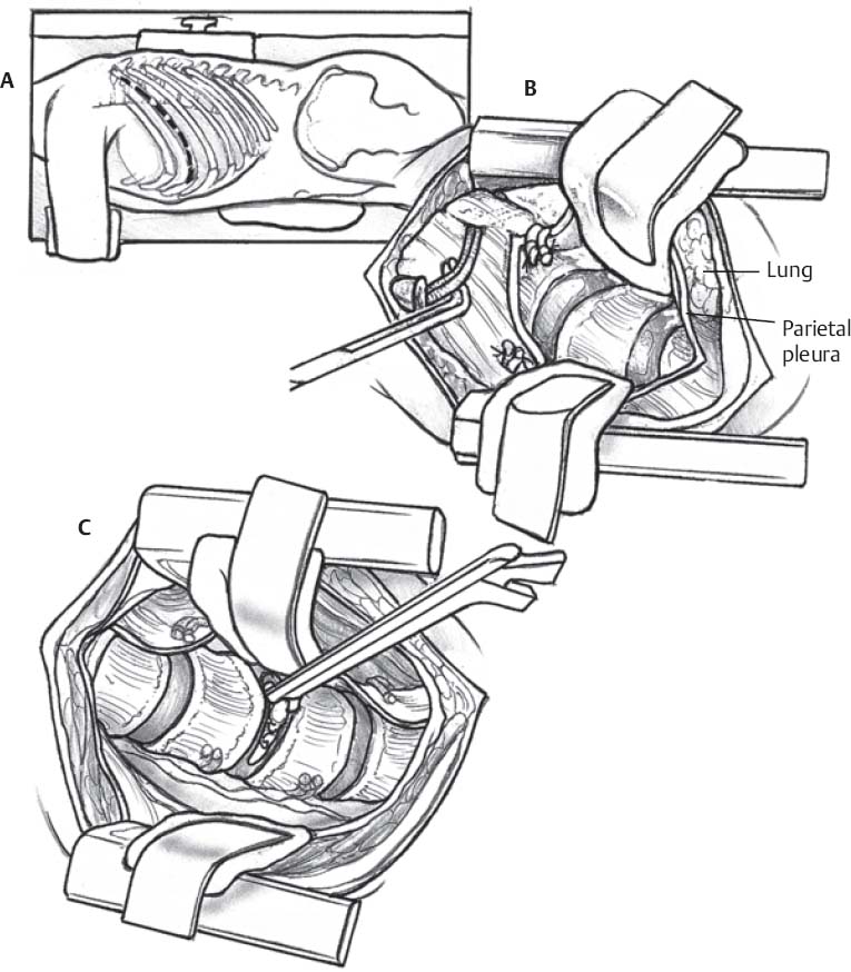
♦ Intraoperative (Fig. 105.1)
Exposure
- Access surgeon makes for a good “team” experience
- Remove rib 1 to 2 levels above target level (remove 6th rib to access T7–T8 or T8–T9)
- Resection of a rib yields bone graft material and reduces the extent of rib retraction needed for a given exposure
- Loupe magnification is generally adequate
- Multiple segmental vessels may be divided as needed on the same side, even on the left
- The sympathetic chain may be divided
- Rib heads may be removed to allow visualization of the transverse processes and pedicles
- Obtain sufficient fluoroscopic and/or plain film images until extent of exposure and level is confirmed
Decompression
- Decompression via standard techniques (long instruments and/or drill attachments may be helpful)
- Consider spinal drainage if dura opened or cerebrospinal fluid (CSF) noted
< div class='tao-gold-member'>
Only gold members can continue reading. Log In or Register to continue
Stay updated, free articles. Join our Telegram channel

Full access? Get Clinical Tree


