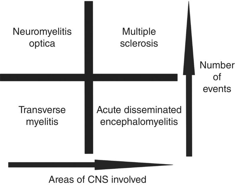
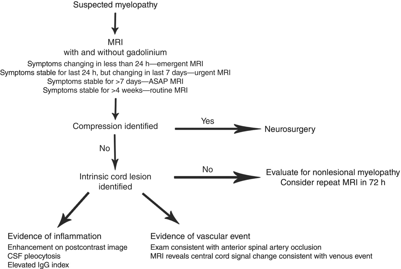
14 Benjamin M. Greenberg Department of Neurology and Neurotherapeutics and Department of Pediatrics, University of Texas Southwestern, Dallas, TX, USA Transverse myelitis (TM) and acute disseminated encephalomyelitis (ADEM) are immune-mediated conditions that can affect the central nervous system (CNS). While classically described as demyelinating disorders, some patients may also have damage to the underlying axons leading to long-term symptoms. The semantics and nomenclature related to these conditions have been the source of much confusion, and large-scale controlled trials have been lacking. This chapter will address issues related to nomenclature, biology, clinical presentation, clinical evaluations, and treatment issues related to these rare but important conditions. First, relative to nomenclature, demyelinating disease terminology has been a source of confusion for patients, families, and practitioners. For example, TM is defined as inflammation of the spinal cord. When it occurs in isolation, it is considered primary or idiopathic TM. Yet, in some patients, TM can be part of ADEM (when both the brain and spinal cord are affected), or it can occur as a relapse in multiple sclerosis (MS) or neuromyelitis optica (NMO) patients. In general, primary TM and ADEM are considered monophasic events, while other conditions are frequently recurrent. Thus, one schematic for considering these conditions is outlined in Figure 14.1. Figure 14.1 Categorization of CNS demyelinating conditions based on temporal course and lesion distribution. The approach to patients with TM and ADEM is quite similar—recognize the condition, treat the inflammation, determine if there is an underlying cause that puts the patient at risk for recurrent events, and manage long-term symptoms. The remainder of this chapter will deal with each condition separately. The term TM was first used in 1948 by an English neurologist, Dr. Suchett-Kaye, to describe a case of rapidly progressive paraparesis with a thoracic sensory level, occurring as a postinfectious complication of pneumonia. While the term myelitis had been used in the 1800s to describe a variety of myelopathic events, transverse was added to recognize the commonality of sensory changes in a banding pattern. There are approximately 1500 cases of idiopathic TM per year, but no large-scale epidemiology studies have been completed (Bhat et al. 2010). Studies that include TM events that are possible first events of MS suggest an incidence of 24.6 cases per million population or approximately 8100 cases per year in the USA (Young et al. 2009). While primary (idiopathic) TM is more common in children than adults, it can occur at any age. Pathologically, patients with TM have evidence of inflammation and demyelination within the white matter of the spinal cord. While there are various publications outlining the pathology associated with secondary TM (lupus-associated TM, NMO-associated TM, MS-associated TM, and sarcoid-associated TM), there are few publications of the pathologic findings seen in idiopathic TM from biopsy or autopsy material (Nakano & Ogashiwa 1995; Pittock & Lucchinetti 2006). The majority of evidence suggests a lymphocytic infiltrate enters the spinal cord and coordinates an attack that can lead to abrogation of signal transmission, demyelination, axonal injury, and neuronal cell body injury. While the majority of adult TM cases involve white matter changes in the spinal cord, pediatric patients are increasingly being identified with mixed white matter and anterior horn cell damage (Howell et al. 2007). Often, because a condition is rare, its rarity is used to justify delays in diagnosis. Yet, in the case of TM, recognition of the condition starts with recognition of a myelopathy. Evidence of spinal cord dysfunction, regardless of cause, should be recognized as a potential neurologic emergency. The most common presenting symptoms of a myelopathy include weakness, loss of sensation, pain, bowel or bladder dysfunction, and/or ataxia. A careful history and physical assessment are necessary in order to accurately localize a lesion to the spinal cord. Patients who present with a sensory level or acute urinary retention should be considered to have a spinal cord disorder until proven otherwise. Numerous patients have been sent home from offices or emergency rooms in the early stages of myelitis, only to return with severe weakness that could have been treated earlier. The most common misdiagnoses for myelopathy and, hence, TM include neuropathy (i.e., Guillain–Barré syndrome), pinched nerves, urinary tract infections (in the setting of acute urinary retention), and stroke. Careful consideration of possible spinal cord pathology should be given to all patients presenting with acute or subacute neurologic events. Once a potential myelopathy is clinically recognized, a standard workup should be initiated to determine if the cause is inflammatory (myelitis). In 2002, a working group proposed diagnostic criteria for TM. These recommendations have been incorporated into Figure 14.2, which outlines the initial management of a patient with suspected myelopathy (Transverse Myelitis Consortium Working Group 2002). Of note, there are no published standard of care guidelines for the timing of studies suggested in Figure 14.2, but based on the need for urgent identification of patients, Table 14.1 lists some suggested time considerations. These recommendations are based on the literature supporting emergent surgical intervention for compressive myelopathies and the growing anecdotal evidence supporting early treatment for myelitis (Park et al. 2006). Figure 14.2 Recommended diagnostic pathway for myelopathic patients. Table 14.1 Timing of MRI Based on the Last Time a Patient’s Myelopathic Symptoms Progressed While an MRI is critical for identifying a compressive cause of myelopathy, it is also critical for identifying intrinsic cord pathology categorized as noncompressive. An MRI of the spinal cord (at and above the level of dysfunction) should be obtained with and without gadolinium. The administration of contrast can be useful for determining the etiology of compressive and noncompressive lesions. If a patient does not have a compressive cause of myelopathy, their workup should then include a lumbar puncture for CSF analysis. TM is suspected in patients with myelopathy and evidence of inflammation, defined as gadolinium enhancement, pleocytosis, or an elevated IgG index (Transverse Myelitis Consortium Working Group 2002, August 27). Of note, these parameters have unknown sensitivity and specificity levels, but are useful tools when evaluating patients. While it is possible for a myelitis patient to have no gadolinium enhancement on MRI and a normal CSF, these features should raise suspicion for alternate explanations. Likewise, patients with vascular events of the cord can have postcontrast enhancement on MRI and abnormal CSF analyses, so the tests are not specific for myelitis (Morimoto et al. 1996). Finally, it is worth noting that an isolated elevated CSF protein is the least specific test in myelopathy patients as it can be abnormal in most CNS disorders making its significance difficult to interpret. Once acute therapy is initiated, practitioners should pursue a workup to try and identify the underlying cause of spinal cord inflammation. The majority of secondary myelitis cases are caused by MS, but NMO, sarcoidosis, systemic lupus erythematosus (SLE), Sjögren syndrome, antiphospholipid syndrome, and copper deficiency should be considered. A typical workup is summarized in Table 14.2. Table 14.2 Standard Evaluation of TM Patients Once a patient is suspected of having TM, a healthcare provider is responsible for initiating therapy quickly while continuing to evaluate the patient for underlying causes. The historical standard of care for acute and subacute myelitis is the use of high-dose corticosteroids. There are few, if any, contraindications to high-dose corticosteroids; thus, practitioners should feel comfortable giving doses while evaluations are in progress.
Transverse Myelitis and Acute Disseminated Encephalomyelitis
Introduction

Transverse myelitis
Epidemiology
Pathobiology
Clinical presentation
Differential diagnosis and diagnostic criteria

Timing of Symptom Evolution
Timing of MRI
Less than 24 h
STAT (within hours)
24 h to 7 days
Urgent (same day)
7–30 days
Within 48 h
More than 30 days
Next available
Test
Reason
Brain MRI
Multifocal brain lesions suggest MS
NMO-IgG
NMO
SSA/SSB
Sjögren-associated TM
ANA
Lupus-associated TM
Anticardiolipin antibody
Antiphospholipid syndrome-associated TM
Copper
Copper deficiency-associated myelopathy
Vitamin B12
Subacute combined degeneration
RPR
Tabes dorsalis
Chest CT scan
Sarcoidosis
CSF oligoclonal bands
Associated with MS
Acute treatment
Stay updated, free articles. Join our Telegram channel

Full access? Get Clinical Tree


