Treatment of Cervical Spine Instability in the Pediatric Patient


Treatment of Cervical Spine Instability in the Pediatric Patient


Denis S. Drummond

Stephanie R. Cody

Nina Agrawal

John P. Dormans


Instability of the cervical spine in children can result from a large spectrum of conditions with a wide range of congenital and acquired etiologies. This chapter focuses on the presentation, evaluation, and treatment of cervical spine instability occurring in children following trauma or in association with congenital anomalies of the cervical spine. Table 37.1 lists and categorizes the most frequently encountered causes of cervical instability.

The cervical spine in children can be divided into the upper cervical spine, extending from the occiput to the C2-C3 disc space, and the lower cervical spine, which extends caudally from C3. Compared to the lower cervical spine, the upper cervical spine is unique in its embryology, developmental anatomy, and biomechanics. At the lower levels, development occurs similarly to the thoracic and lumbar spine (1). In contrast, development at the upper levels, particularly the atlas and axis, is unlike development in any other part of the spine. These distinct developmental features can determine the site and severity of the injuries. For example, the most serious cervical injuries observed in children, particularly those resulting in neurologic injury, occur in the upper cervical spine. This is likely the result of the unique anatomy and biomechanics occurring there. Also, the pediatric spine is continually changing with growth, maturation, and ongoing ossification of vertebral bodies as observed on radiographs. Particularly, during the first decade, the polar growth centers adjacent to the vertebral end plates and the many synchondroses present in the immature skeleton demarcate likely sites of vertebral injury. For example, the basal synchondrosis caudal to the base of the dens is the site of fracture separation in younger children (2). This susceptibility to injury is covered more completely in the biomechanics section. Besides affecting stability, changes of the immature spine during growth make the radiographic evaluation of children difficult following injury or when presenting with congenital vertebral anomalies. This is particularly a problem for those less familiar with the radiographic appearance of the normal pediatric cervical spine.
BIOMECHANICS

The mechanics of the spine in children differs from that of healthy adults. The facet joints tend to be shallower and are less competent in the immature spine. Furthermore, the comparative ligamentous laxity of children also has adverse effects on stability. Third, the atlantooccipital and atlantoaxial joints are less well supported and tend to be less stable in children. Finally, with high-energy injuries, the relatively large head of young children magnifies the acceleration forces on the upper cervical spine. Accordingly, when subjected to acceleration and deceleration forces, younger children are particularly vulnerable to injury in the upper cervical spine. Specifically, the spinal cord is at risk for serious traction injury. With failure of the supporting soft tissues, the stability of the discs, facet joints, and synchondroses may be exposed to shear forces. Although the pediatric spine can tolerate subluxation and distraction, the spinal cord cannot. With stretch beyond tolerance, myelopathy will occur, a phenomenon known as spinal cord injury without radiographic abnormality, or SCIWORA.


DEFINITIONS OF INSTABILITY

The range of values used to define normal stability is slightly broader for children than for adults. For example, the upper limit of “normal” for translation at the atlantodens interval (ADI) (C1-C2) is 3 mm for adults and 4 mm for children. Further, an acceptable increase in translation is dependent not only on the measurement but also on the space available for the cord (SAC) as observed on magnetic resonance images (MRI). A good rule of thumb is that one-third of the space of the cross-sectional diameter can be available for the dens, one-third for the spinal cord, and one-third is needed as available space (3). A borderline measurement for increased translation is better accepted in a capacious canal rather than in a smaller, or stenotic, one. The methods of measurement and the established normal values when observed on lateral flexion and extension films are defined below.


TABLE 37.1 Classification of Cervical Instability


Causes


Subtypes


Congenital


  • Vertebral (bony anomalies)


    a. Craniooccipital defects (occipital vertebrae, basilar impression, occipital dysplasias, condylar hypoplasia, occipitalized atlas)


    b. Atlantoaxial defects (aplasia of atlas arch, aplasia of odontoid process)


    c. Subaxial anomalies (failure of segmentation and/or fusion, spinal bifida, spondylolisthesis)


  • Ligamentous or


  • Combined anomalies found at birth as an element of somatogenic aberration


Syndromic disorders (i.e., Down syndrome, Klippel-Feil syndrome, 22q11.2 deletion syndrome, Marfan’s syndrome, Ehlers-Danlos syndrome)


Acquired


Trauma


Infection (pyogenic/granulomatous)


Tumor


Inflammatory conditions (i.e., juvenile rheumatoid arthritis)


Osteochondrodysplasias (i.e., achondroplasia, spondyloepiphyseal dysplasia)


Storage disorders (i.e., mucopolysaccharidoses)


Miscellaneous (i.e., postsurgery)


From Hosalkar HS, Agrawal N, Drummond DS. Congenital osseous anomalies of the cervical spine. In: Bridwell KH, ed. The Textbook of spinal surgery, Philadelphia: Lippencott Williams and Wilkins, 2011:1199.


ATLANTOOCCIPITAL MOTION

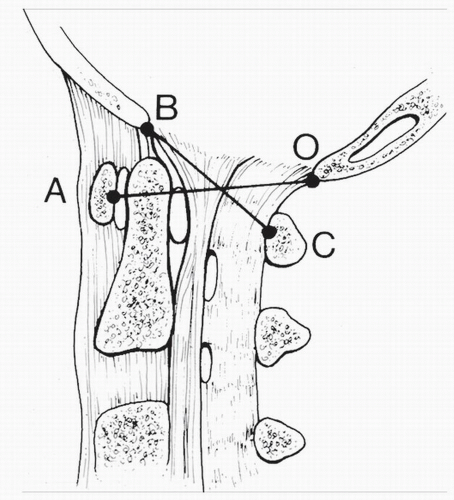
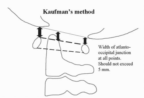
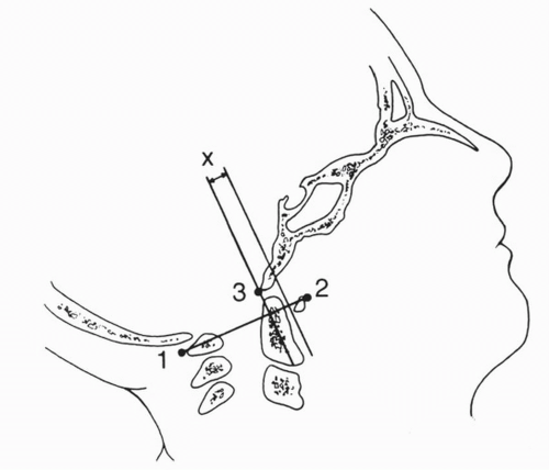
Excess atlantooccipital translation causing instability is best observed on the lateral extension radiograph. The three commonly used parameters for stability at this level are the Power’s ratio (Fig. 37.1), the Harris technique, and the Kaufmann technique. The Power’s ratio and Harris technique both measure horizontal translation, whereas the Kaufmann technique (Fig. 37.2) measures vertical displacement. Horizontal translation should not exceed 1 mm in the adult and 2 mm in children (4 mm is accepted in patients with Down syndrome). Vertical displacement should not exceed 5 mm. The authors prefer the Weisel-Rothman method of measuring horizontal translation (Fig. 37.3) because it is simple and offers a clear view of the skeletal end points. The measurement extends from the anterior edge of the foramen magnum (basion) to the posterior surface of the anterior atlas. These are usually measured and compared in both the neutral position and maximal hyperextension (4).


ATLANTOAXIAL MOTION

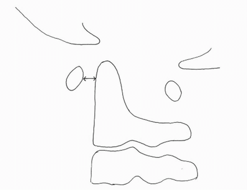
Atlantoaxial instability or excess translation is best observed on lateral radiographs done in flexion and compared with the lateral neutral view. The ADI is the accepted and standard measurement (Fig. 37.4). In adults, this should not exceed 3 mm, and in children 4 or 5 mm is usually accepted as the upper limit of normal for this index.


Figure 37.1. Atlantooccipital subluxation is determined using Power’s ratio: for patients without subluxation, BC ≤ AO ≤ 1. (From Seminars in spine surgery, pediatric cervical instability: diagnosis and treatment concepts, vol. 8, No 4, Philadelphia: W.B. Saunders Company, 1996).


SUBAXIAL MOTION

There is no accepted norm for subaxial translation to define instability in children; we have arbitrarily defined the norm as 2 mm. Subaxial instability may be caused by trauma and ligamentous laxity. One typical site of injury is the cervicothoracic junction (5). Subaxial translation is best appreciated on the lateral flexion views. Again, it is important to emphasize that the SAC is an important issue when evaluating for safety of segmental translation occurring distal to the axis.


Figure 37.2. The Kaufman technique is useful for determining vertical displacement on flexion-extension films.


Figure 37.3. Wiesel-Rothman method for measuring horizontal translation. A line is drawn from the caudal tip of the posterior arch of the atlas (1) to the center of the anterior arch of the atlas (2) and intersected by a line drawn through point 3 at the inferior tip of the basion. A second line is drawn parallel to the line through point 3 at the anterior arch. Horizontal translation is measured as the distance between the two parallel lines (x). (From Hosalkar HS, Drummond DS. Pediatric cervical spine. Orthopaedic knowledge update 3: SPINE. Section Editor Drummond DS. 3rd ed. 2006.


DIAGNOSING INSTABILITY

Performing a clinical examination to determine instability in pediatric patients is difficult compared to doing so in adults. One reason for this difficulty is that children, particularly young ones, have trouble cooperating with the examination. When evaluating trauma in the young patient, one frequently needs a high index of suspicion for serious cervical injury. It is important to seek clues that indirectly suggest cervical trauma, such as a head or facial injury, seatbelt injury, or an unconscious patient. All of these observations should raise suspicion of an associated cervical injury. Cervical examination should include looking for tenderness, swelling, and bruising. A finding of torticollis after trauma may indicate a C1-C2 rotatory subluxation or a minor spinal cord injury. Spasm of the sternocleidomastoid muscle may be suggestive of spinal injury. The “seat belt sign,” a transverse patch of skin contusion across the abdomen, should alert the clinician to associated injuries to the abdominal viscera, the cervical spine, and the lumbar spine. In addition, a careful neurologic examination, including assessment of the bulbocavernosus reflex, should be performed.


Figure 37.4. Diagrammatic illustration showing the ADI (arrow).


RADIOGRAPHIC EXAMINATION TO DETERMINE STABILITY

Because of the unique features associated with skeletal immaturity, the pediatric cervical spine poses a difficult radiographic study for most clinicians, particularly those less experienced in evaluating the immature spine. Table 37.2 lists the radiographic features that can be observed in the normal pediatric spine. At times these observations are mistakenly interpreted as spinal pathology. Similarly, instability is frequently overdiagnosed in the young because the incomplete ossified vertebrae give a false impression of translation or pseudosubluxation.
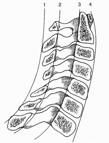
By applying the sublaminar line shown in Figure 37.5, a correct determination of stability may be made. Misdiagnosis may also occur in the case of an actively crying child. Crying can cause the prepharyngeal space to appear enlarged on the lateral radiograph, erroneously suggesting traumatic edema or hematoma.


TABLE 37.2 Unique Features of the Normal Pediatric Spine


Unique features of the pediatric spine


General features


Secondary centers of ossification of the spinous processes may mimic fractures


Rounding of anterior vertebral body may give the appearance of a wedge compression fracture


Horizontal facets and ligamentous laxity allow greater intersegment mobility


Decreased cervical lordosis


Wider prevertebral soft tissues may mimic swelling


Special features


C1 multiple ossification centers may mimi fractures


Absent ossification of anterior arch of C1 may be interpreted as C1-C2 instability


C1-C2 atlanto-dens interval may be up to 4.5 mm in normal children


C2 normal posterior angulation of odontoid (4% of children) may mimic fracture


Ossiculum terminale may be confused with a fracture


Basilar synchondrosis may be confused with a fracture


C2-C3 and C3-C4 pseudosubluxation can be mistaken for instability


From Hosalkar HS, Agrawal N, Drummond DS. Congenital osseous anomalies of the cervical spine. In: Bridwell KH, ed. The textbook of spinal surgery, Philadelphia: Lippencott Williams and Wilkins, 2011:1198.


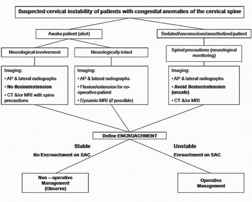
Although lateral flexion-extension radiographs are considered the gold standard for stability when evaluating the cervical spine, this study is unsafe for the unconscious patient or uncooperative young child. Because of relative ligamentous laxity in children, the cervical spine can deform more than the spinal cord can tolerate; SCIWORA may thus occur. If spontaneous reduction occurs, the unwary observer may believe that the spine is structurally normal and then fail to provide the appropriate stability and environment needed to prevent further spinal cord injury. For this reason, dynamic radiographic studies done to confirm stability, that is, lateral and flexion series, are unsafe and therefore contraindicated in unconscious or uncooperative young patients. In contrast, advanced imaging, such as MRI and computed tomography (CT) scanning, is appropriate in these cases. Frequently, MRI will reveal a high signal, particularly in the T2-weighted images. A high signal within bone suggests an occult fracture; in the supporting soft tissues, it implies injury that could compromise the stability of the spine. MRI can also assist with diagnosis of injury to the spinal cord. A CT is useful for demonstrating the exact location of the occipital condyles and can offer a sagittal reconstruction. Dynamic CT may help elaborate the nature of instability and document fixed subluxation in patients with atlas ring fractures or atlantoaxial rotary subluxation. Figure 37.6 presents an algorithm showing the appropriate management of traumatic cervical instability in both the conscious and unconscious patient.


Figure 37.5. Illustration showing the normal relationships in the lateral cervical spine. The spinolaminar line (3) is helpful for evaluating stability in very young patients. (From Copley LA, Dormans JP. Cervical spine disorders in infants and children. J Am Acad Orthop Surg 1998;6(4):204-214, p. 207, Fig. 5.)


Figure 37.6. Preferred treatment algorithm for cervical instability dividing patients into conscious and unconscious. (Modified from Hosalkar HS, Agrawal N, Drummond DS. Congenital osseous anomalies of the cervical spine. In: Bridwell KH, ed. The textbook of spinal surgery, Philadelphia: Lippencott Williams and Wilkins, 2011:1199.)


TRAUMATIC CERVICAL INSTABILITY

Cervical spine injury occurs in 1% of all pediatric trauma cases (6,7). As described above, these injuries are more often found in the upper cervical spine of the young child and are more likely to be associated with neurologic involvement than injuries occurring distal to that area (8). Thus, the fatality rate for young children is double that found in adults (6,7,9). Some of the common injuries of the cervical spine are outlined in Table 37.3.


ATLANTOOCCIPITAL DISLOCATION

Atlantooccipital dislocation is a rare but frequently fatal injury. Significant disruption is more common in pediatric than adult injuries because of the relatively large size of the cranium in younger children. Hyperextension, lateral rotation, or forward flexion all may be the mechanism of injury. Almost 20% of patients with atlantooccipital instability are free of major neurologic injuries, although some may experience cranial nerve injury, vomiting, headache, torticollis, or minor weakness (10, 11 and 12). The diagnosis may be masked or overlooked in patients with concomitant brain injury. One should be alert for clues to cord injury, such as spinal shock and urinary retention in cases of complete cervical lesions as well as neck pain, tingling, or spasticity out of proportion to cognitive impairment in partial lesions (13).

In spite of the high fatality rate associated with atlantooccipital dislocation, in a study from our center, we observed that 5 of 16 children with this injury survived following timely and aggressive management, as described in the treatment section. Further, we established a clue to prognosis for those patients with the best outlook for survival and significant improvement. We observed that 4 of the 16 patients were able to phonate or cry at the time of the emergent provision of the tracheal airway. We call this the “moan” sign; it indicates incomplete brainstem injury and some preservation of the nerves that support spontaneous ventilation. All four of the patients that could moan or phonate at the time of intubation regained considerable neurologic function (14). In patients with atlantooccipital dislocation, it is important to provide a timely reduction and stability for the spine with an appropriately applied halo vest, get an airway established, begin a steroid protocol to control spinal cord edema, and stabilize the patient systemically to prepare for surgical arthrodesis.


TABLE 37.3 Common Cervical Spine Injuries in Children


Atlantooccipital trauma


  • Jefferson fracture


  • Atlantooccipital dislocation


  • Odontoid fracture


Atlantoaxial trauma


  • Hangman’s fracture


  • Traumatic spondylolisthesis


  • Atlantoaxial subluxation


  • Atlantoaxial rotational dislocation


Subaxial trauma


  • Traumatic subluxation with or without fracture


  • Traumatic fracture dislocations


  • Anterior/posterior/lateral element fractures in isolation or combination


SCIWORA


JEFFERSON FRACTURE

Fractures of the atlas, or Jefferson fractures, are also rare in children and are best observed on axial images of the CT scan. Often, when an atlas fracture does occur, only one may be seen opposite a synchondrosis that is plastically deformed. This can give the erroneous impression that the injury was not high energy or severe. The clinician, therefore, should be alert to other signs. For example, a widening of the lateral masses greater than 7 mm suggests that the transverse ligament is ruptured, and this is indeed an urgent indication for immobilization, usually in a halo vest. With a more stable situation and no significant widening of the lateral masses, an orthotic collar may suffice (15).


ODONTOID FRACTURE

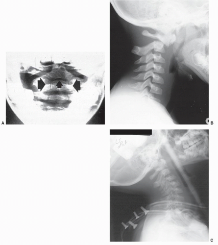
Fractures of the odontoid are the most common fractures of the cervical spine in children that may be associated with instability. They occur more frequently in younger children and most often involve the basilar synchondrosis, which is situated just distal to the base of the dens (Fig. 37.7). In a recent study, we observed 23 children with odontoid fractures (2). The observations derived from the study validate our treatment principles. First, we found that advanced imaging was frequently required to define the injury absolutely. Without advanced imaging data, diagnosis may be delayed, running the risk of more serious injury. Seventeen of the 26 patients studied had a fracture through the basilar synchondrosis. All displaced fractures through the synchondrosis were displaced ventrally and reduced with a gentle extension followed by immobilization. Successful gentle reduction depends on the anterior periosteal hinge of the odontoid being intact and providing a buttress and stability during the extension reduction. The spine was then immobilized by a Minerva cast (x patients) or halo vest (y patients) for 6 to 8 weeks, depending on age. Of the 17 patients in our series with fracture through the synchondrosis, one did not survive an associated head injury and another was lost to follow-up. Accordingly, we were able to follow 15 patients. Thirteen of the 15 healed in a halo vest after a gentle reduction in extension, while two required an early arthrodesis because of serious associated injuries.

For patients with open synchondroses distal to the base of the odontoid, we developed a new classification system, which is outlined in Figures 37.8 and 37.9. For the six patients with closed synchondroses, we found that the Anderson and D’Alonzo (16) classification developed for the adult population was appropriate. Further, the treatment principles for these patients are similar to those advised for adults.


Figure 37.7. Dens fractures through the basilar synchondrosis are the most common cause of traumatic cervical spine instability in children. Radiographs courtesy of Denis Drummond, MD.


HANGMAN’S FRACTURE

Hangman’s fractures with spondylolisthesis at C2 are rare in children but should remain in the differential diagnosis following significant trauma, particularly when associated with a seatbelt injury. This fracture is best diagnosed on CT scans with sagittal and frontal formatting.

Jun 29, 2016 | Posted by in NEUROLOGY | Comments Off on Treatment of Cervical Spine Instability in the Pediatric Patient

Full access? Get Clinical Tree

Get Clinical Tree app for offline access