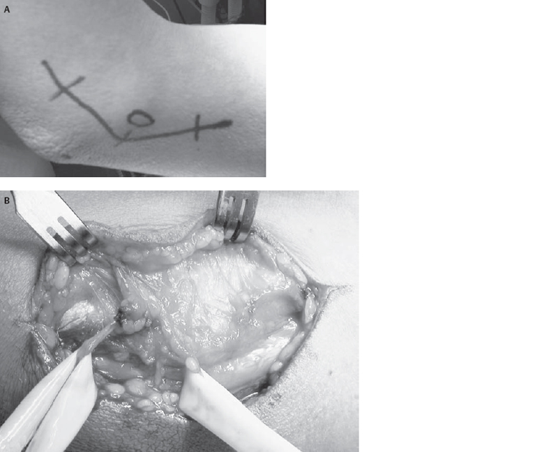
27 Ulnar Nerve Entrapment at the Elbow A 66-year-old, right-handed man presented with progressive right-hand numbness and weakness. He had had a successful left carpal tunnel release many years prior and was not known to be diabetic. His recent symptoms started about 1 month after removal of his right parotid adenocarcinoma, which also required subsequent radiotherapy. He initially noticed paresthesias and decreased fourth and fifth digit sensation while using the mouse on his computer. Concomitantly, there was significant pain and tenderness over the medial elbow region. Over the next 3 months he lost his hand muscles’ bulk and strength with decreased gripping ability. His neurological examination revealed profound weakness of the flexor digitorum profundus (FDP) of the fourth and fifth fingers (grade 3) with marked weakness and atrophy of hand intrinsics. Both Froment and Wartenberg signs were positive. The sensation to pinprick was decreased in the little and ulnar aspect of the ring fingers. There was no Tinel phenomenon overlying the ulnar nerve in the cubital tunnel. The electrophysiological study demonstrated denervation of ulnarinnervated muscle groups. A localized nerve conduction block could not be demonstrated because of severe axonal loss. The clinical and electrophysiological findings were consistent with severe compressive ulnar neuropathy at the elbow with advanced motor signs. Intraoperative exploration of the ulnar nerve in the cubital tunnel revealed focal compression just deep to the arcuate ligament and common aponeurosis of the flexor carpi ulnaris (FCU) muscles. The nerve was decompressed by widely incising all soft tissue compressive elements, and it was elected not to transpose the nerve. His long-term follow-up confirmed complete resolution of pain with substantial sensory improvement and slight motor recovery. Hand dexterity was improved considerably. Ulnar nerve entrapment at the elbow The ulnar nerve anatomy in the arm, elbow, and forearm is relatively constant. It is the terminal continuation of the medial cord that has components of C7, C8, and T1 roots. Initially, it courses between the axillary artery and vein, just anterior to the subscapularis muscle and posterior to the inferior fibers of the pectoralis minor. It courses into the upper medial arm anterior to the latissimus dorsi tendon in a groove between the coracobrachialis muscle laterally and the long and medial head of the triceps brachii muscle posteriorly. At about the middle of the arm, the ulnar nerve (joined by the superior ulnar collateral artery) passes through the upper portion of the medial inter-muscular septum to descend on the anterior aspect of the medial head of the triceps or within the triceps in 25% of cases. The connective tissue barrier, which is traversed as the ulnar nerve descends from the anterior into the posterior compartment, is thickened at ˜8 cm above the medial humeral epicondyle. This thickened fibrofascial structure, called the arcade of Struthers, is present in ˜70% of the population. Although it is not a common site for entrapment, it may take importance as a secondary site for ulnar nerve kinking after anterior transposition without complete release of this structure. In the elbow the nerve lies in the postcondylar groove on the dorsum of the humeral (medial) epicondyle. The articular branches to the elbow joint are given off the ulnar nerve at this level before entering into the cubital tunnel between the medial epicondyle and olecranon. In the cubital tunnel, the ulnar nerve is covered by the arcuate ligament, which extends from the medial humeral epicondyle (lateral wall) to the tip of the olecranon (medial wall). The elbow joint capsule and the medial collateral ligament form the floor of the cubital tunnel. The taut, variably thick arcuate ligament with its sharp edge, which merges with the common aponeurosis of the FCU, is the common site for ulnar nerve constriction. The rare presence of the ulnar-innervated anconeus epitrochlearis muscle, which also extends from the medial epicondyle to the olecranon, can also contribute to ulnar nerve entrapment in the cubital tunnel. The first muscular branch of the ulnar nerve is to the FCU. There are up to four branches coming off the ulnar nerve from 4 cm above to 10 cm below the medial epicondyle. The ulnar nerve subsequently enters the forearm between the humeral and ulnar head of the FCU and descends over the medial side of the forearm and FDP covered by the FCU. The internal topography of the ulnar nerve fascicles in the elbow region with preferential deep location of FCU and FDP fascicles explains their sparing in ulnar nerve entrapment at the elbow level. The posterior branch of the medial cutaneous nerve of the forearm crosses the ulnar nerve in the subcutaneous plane from 6 cm proximal to 4 cm distal to the medial epicondyle. Preservation of this nerve during ulnar nerve exposure is important to avoid numbness or painful neuroma formation (Fig. 27–1). The capacity of the cubital tunnel is dynamic and will decrease up to 55% with the combination of elbow and wrist ulnar flexion. This is due to stretching and tautness of the arcuate ligament in flexion, and active FCU contraction. The presence of a shallower groove on the inferior surface of the medial epicondyle as opposed to the posterior surface will raise the floor of the cubital tunnel during flexion. Fusiform enlargement of the ulnar nerve behind the medial epicondyle has been seen in ˜50% of examined normal cadaveric ulnar nerves and is thought to be due to an increase in the nerve connective tissue. This focal enlargement could potentially aggravate compression in the cubital tunnel, especially with upward and downward sliding of the ulnar nerve during elbow movement. The ulnar nerve slides ˜10 mm proximally and 3 mm distally around the elbow during the flexion-extension maneuver. The pathophysiology of ulnar entrapment therefore seems to be due to a combination of compression, stretch, and motion of the ulnar nerve about the elbow joint.
 Case Presentation
 Diagnosis
 Anatomy

Stay updated, free articles. Join our Telegram channel

Full access? Get Clinical Tree


