Fig. 48.1
ADEM related to vaccination. Axial FLAIR images (a–f) obtained approximately 2 weeks after the administration of the vaccine demonstrate numerous hyperintense foci (arrows) involving the infratentorial and supratentorial white matter and thalami. Selected FLAIR images (g–i) obtained approximately 1 month later show interval resolution of the lesions
Certain vaccines can also result in Guillain-Barre syndrome. Patients present with acute flaccid paralysis, as well as sensory abnormalities and autonomic dysfunction. Salient features of Guillain-Barre syndrome on MRI include diffuse, smooth mild thickening and enhancement of the cauda equina nerve roots, preferentially the anterior nerve roots (Fig. 48.2). The spinal cord can also be affected, demonstrating patchy areas of high T2 signal and variable degrees of enhancement. There may be concomitant brain involvement; therefore, the brain should be imaged as well in these cases. Ultimately, the diagnosis is based on the temporal relation to vaccination and exclusion of other potential etiologies.
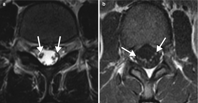
Fig. 48.2
Guillain-Barre disease related to vaccination. Axial T2-weighted (a) and post-contrast T1 (b) MR images show preferential thickening and enhancement of the anterior cauda equina nerve roots (arrows)
48.4 Differential Diagnosis
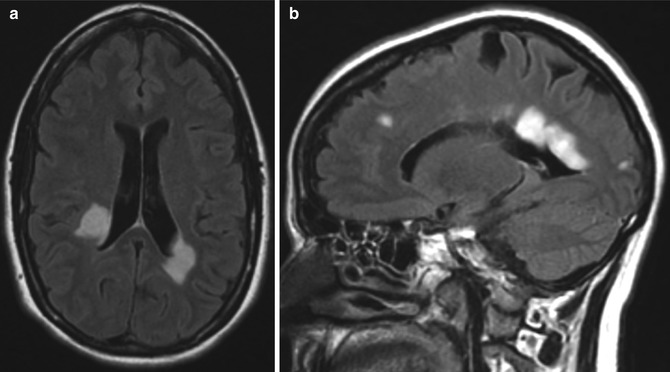
The main differential diagnoses for vaccine-induced ADEM include ADEM caused by other etiologies, other demyelinating conditions such as multiple sclerosis, and viral encephalitis. Postinfectious ADEM is essentially indistinguishable from postimmunization ADEM on imaging, and clinical history may suggest the precipitating factor. Although multiple sclerosis may resemble ADEM on MRI, multiple sclerosis is a progressive disease rather than a monophasic one. Nevertheless, findings on conventional MRI that favor multiple sclerosis over ADEM include the absence of a diffuse bilateral lesion pattern, presence of black holes, and presence of two or more periventricular lesions (Fig. 48.3). In addition, magnetization transfer sequences can help distinguish ADEM from MS, in that normal-appearing brain on T2-weighted images has normal magnetization transfer ratio, whereas in multiple sclerosis measurements are decreased in both areas. It is important, but not always straightforward to differentiate viral encephalitis from ADEM. On MRI, viral encephalitis typically demonstrates bilateral diffuse areas of T2 hyperintensity involving the cerebral cortex and the underlying white matter and, to a lesser extent, the basal ganglia, brainstem, and cerebellum (refer to Chap. 3). However, the particular distribution of the lesions may depend upon the particular viral agent. Associated enhancement may or may not be present, but tends to be leptomeningeal when present.