Cervical Deformity
Matthew Grosso
Michael Steinmetz
Edward Benzel
The normal lordotic curvature of the cervical spine is critical to maintaining sagittal alignment and spinal balance. Forces, both internal and external, can affect this alignment and lead to symptomatic conditions that may require clinical and/or surgical intervention. Deformity of the cervical spine is most commonly found in the sagittal plane, presenting as cervical kyphosis. Less common deformities of the sagittal plane include hyperlordosis and mixed swan neck. Changes in the coronal alignment, such as scoliosis, are much less common in the cervical spine than in the lumbar and thoracic regions (1).
The reversal of normal cervical curvature, as seen in kyphosis, can occur through a variety of mechanisms. Possible etiologies include degenerative disease, trauma, neoplastic disease, systemic arthritides such as ankylosing spondylitis (AS) and rheumatoid arthritis, neuromuscular, congenital anomalies, and iatrogenic (postsurgical, postradiation, etc.) (2). Symptomatic patients often present with mechanical neck pain as well as a variety of neurologic disorders related to myelopathy and radiculopathy. For patients with severe cervical kyphosis, chin-on-chest deformity can develop, which limits the ability to perform basic functions including forward gaze, swallowing, and even respiration. Surgical intervention may be warranted for patients with sufficiently symptomatic deformities.
ANATOMY AND BIOMECHANICS
The cervical spine consists of seven vertebrae that extend from the base of the skull (C1) to the upper thoracic cavity (C7). Cervical lordosis is created and maintained by a number of structures. These include the vertebral bodies and intervertebral disks, facet joints, and the supporting ligaments and muscles. Moving superior to inferior through the subaxial vertebrae, there is an increase in vertebral body size, with an exception seen at the C5 to C6 transition (3, 4 and 5). Between the bulky vertebral bodies are the cartilaginous disks that play an important role in vertebral movement and force dispersion. Greater ventral than dorsal disk height contributes to the overall lordosis (6). In the neutral position of a lordotic spine, 36% of a compressive load is distributed among the vertebral bodies and disks along the ventral side of the spine (7). The remaining 64% is supported by the dorsal spinal column. Critical dorsal structures for lordotic integrity include the lamina and facet joints, ligamenta flava, and interspinous ligaments (8,9). Studies investigating damage to these structures have demonstrated their importance in maintaining cervical stability and preventing kyphosis. In examining patients following cervical laminectomy, Saito et al. (8) attributed deformity to resection of one or more spinous processes and/or dorsal ligaments including the ligamentum flavum, supraspinous, and interspinous ligaments. Maeda et al. (10), in examination of patients who underwent laminoplasty, concluded that the dynamic factors of the posterior column (muscles and ligaments) play the most important role in preserving cervical lordosis.
Although a definite range for normal cervical lordosis has never been established, most studies conclude average angles to be between 15 and 25 degrees using perpendicular lines of the dorsal aspects of C2 and C7 (7,11,12). Gore et al. (11) observed a small difference between the lordotic curvature of men and women, with average angles of 16 to 22 degrees and 15 to 25 degrees, respectively. Although it was initially thought that age and disk degeneration would
contribute to kyphosis, Gore’s original study and 10-year follow-up study of 159 asymptomatic patients concluded that aging and degenerative effects lead to an increase in the lordotic angle (11,13). The study noted an increase of 7 to 10 degrees between the third and seventh decades of life.
contribute to kyphosis, Gore’s original study and 10-year follow-up study of 159 asymptomatic patients concluded that aging and degenerative effects lead to an increase in the lordotic angle (11,13). The study noted an increase of 7 to 10 degrees between the third and seventh decades of life.
Lordosis contributes to extension of the spinal column and reduces the length of the spinal canal. Breig first demonstrated the extent of change in spinal cord length that occurs during extension and flexion (14,15). He measured an increase of up to 75 mm in spinal cord length during flexion. In this scenario, the spinal cord is subject to the greatest tensile forces. The natural curvature of the spine, including cervical lordosis, allows the cord to traverse through a shorter course with minimal tensile stresses (4). Increasing kyphosis results in tensile stresses to the spinal cord generated by pulling of the cord against the apex of the deformity. This is known as the sagittal bowstring effect (16) (Fig. 86.1). Spinal cord tension against the apex can also lead to compression of the ventral neural elements against the bony vertebral bodies or protuberant intervertebral disks (17). Spinal cord vasculature can also be affected, causing subsequent neuronal ischemia. For patients with cervical deformity, examination of the spinal cord in the axial projection often demonstrates flattening at the kyphotic apex (18). These stresses on the spinal cord and the spinal cord’s vasculature contribute to the variety of neurologic symptoms observed in patients with cervical kyphosis (14,16,17,19,20).
As mentioned, the cervical spine is a critical component of normal sagittal balance. Lordosis is a compensatory response to thoracic kyphosis and allows for proper positioning of the head over the trunk. Changes in cervical curvature can be secondary to changes in the lumbar and thoracic spine. Hyperlordosis has been noted in patients with increased thoracic kyphosis (21). These compensatory pathologies must be distinguished from primary deformities affecting the cervical spine.
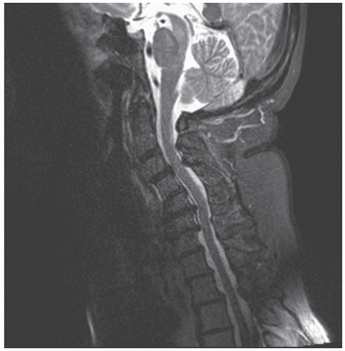 Figure 86.1. Focal kyphosis due to multilevel spondylosis with spinal cord compression at the apex of the deformity. Effective surgical treatment could include direct removal of the source of compression, correction of the kyphosis to eliminate the “bowstring” effect, or a combination of the two. |
No matter the initial cause of cervical deformity, a kyphotic curvature begets further kyphosis. Under normal conditions, the muscles and ligaments of the cervical spine help maintain the lordotic curvature due to the large moment arm relative to the weight-bearing axis. As the degree of kyphosis increases, the head shifts forward, and the distribution of weight favors ventral compression and dorsal tension. An increasingly unfavorable moment arm leads to increasing tensile forces acting on the dorsal ligaments and muscles leading to weakness, fatigue, and failure to support cervical lordosis (17). As the process proceeds, the already compromised muscles are lengthened, further decreasing their efficiency with respect to the length-tension curve of muscular forces.
ETIOLOGIES
POSTSURGICAL KYPHOSIS
Although postsurgical kyphosis can occur following both ventral and dorsal cervical procedures, postlaminectomy kyphosis is the most common iatrogenic cause (2). Destruction of the dorsal tension band and dorsal components of the vertebrae has become a serious concern when performing a laminectomy procedure (Fig. 86.2). The reported prevalence of
kyphotic deformity after cervical laminectomy is quite high, with pediatric patients displaying considerably more risk due to remaining skeletal growth in the face of force imbalance. There is considerable variability in these rates across studies, with values ranging from 37% to 100% (2,22, 23, 24 and 25). In a follow-up of 89 pediatric patients who underwent multiple level laminectomies, Bell et al. (26) found an incidence of cervical kyphosis of 37%, with 15% of patients exhibiting signs of hyperlordosis. An 88% prevalence of postoperative deformity was measured in a study by de Jonge et al. (22), where cervical laminectomy was combined with irradiation treatment of pediatric patients with malignant tumors. Similarly high rates between 88% and 100% have been seen in various other pediatric studies (2,22,23).
kyphotic deformity after cervical laminectomy is quite high, with pediatric patients displaying considerably more risk due to remaining skeletal growth in the face of force imbalance. There is considerable variability in these rates across studies, with values ranging from 37% to 100% (2,22, 23, 24 and 25). In a follow-up of 89 pediatric patients who underwent multiple level laminectomies, Bell et al. (26) found an incidence of cervical kyphosis of 37%, with 15% of patients exhibiting signs of hyperlordosis. An 88% prevalence of postoperative deformity was measured in a study by de Jonge et al. (22), where cervical laminectomy was combined with irradiation treatment of pediatric patients with malignant tumors. Similarly high rates between 88% and 100% have been seen in various other pediatric studies (2,22,23).
 Figure 86.2. Lateral radiograph of a patient with postlaminectomy kyphosis and resultant myelopathic symptoms. The previous surgery involved C4 to C6 laminectomy for a biopsy of an intradural lesion. |
Rates are lower in the adult population, with current estimates varying between 14% and 52%. In a retrospective study of 46 adult patients who underwent laminectomy, Kaptain et al. (27) noted an incidence of 14% of postoperative kyphosis in patients who initially had a lordotic spine, and 30% in those who had a straight spine preoperatively. In a long-term study by Mikawa et al. (28), 64 patients were examined over a 26-month period. Of the original population, 36% had some change in cervical alignment, and 14% had diagnosed kyphosis.
Risk factors for postoperative cervical kyphosis include age, number of laminae removed, preoperative sagittal balance, dorsal tension band stability, presence of intraspinal tumors, irradiation, and extent of laminectomy and facetectomy. Age, as mentioned, is probably the most important risk factor for postoperative deformity. In an effort to remove spinal tumors as a confounding variable when examining incidences in the pediatric population, Yasuoka et al. calculated incidence rates in both adult and pediatric populations that were undergoing laminectomies for conditions that did not cause deformity themselves. The study still found significant differences between the two populations: 46% of the younger population acquired a deformity compared to 6% of the adult population (24). Numerous studies, both clinical and in vitro, highlight the importance of supporting ligamentous and muscular structures, specifically the ligamentum flavum, interspinous ligaments, and dorsal paraspinous muscles (8,10). Increased laxity of dorsal ligamentous structures in the pediatric population is another possible explanation for the increased incidence.
Prophylactic treatment for postlaminectomy kyphosis has developed since its recognition. Facet fusion and lateral mass fixation performed following a laminectomy have shown the ability to reduce rates of postoperative deformity (29,30). Laminoplasty has also significantly reduced the incidence of postsurgical kyphosis (30, 31 and 32). Matsunaga et al. compared outcomes of 64 patients who underwent cervical laminoplasty and 37 patients who underwent cervical laminectomy. They reported a significantly lower incidence of postoperative kyphosis in those who underwent the laminoplasty (31). The incidence of kyphosis following laminoplasty is unclear, but some studies suggest that it is still a concern (33, 34, 35 and 36).
DEGENERATIVE
Cervical kyphosis secondary to degenerative changes is the most common etiology among the elderly. Degenerative kyphosis goes hand in hand with cervical spondylotic myelopathy, the most common spinal disorder among persons greater than 55 years (37). Although Gore et al. (11) concluded that degenerative changes associated with the normal aging process lead to an increase in the lordotic angle, kyphotic deformity is possible for a portion of the population (11). The primary cause of degenerative cervical kyphosis is attributed to changes in disk anatomy. In the nondeformed patient, the height of the cervical disks contributes to the lordotic curve and makes up 15% of the total cervical height (38).
Degeneration of the disks decreases disk height and shortens the anterior column, which leads to a straightening of the cervical spine and possible kyphosis. The contribution of intervertebral disks to the normal lordotic curve is highlighted in the high complication rate of local kyphosis in anterior cervical discectomy (39). Additionally, total disk replacement has shown efficacy in improving sagittal alignment (40, 41 and 42). In a recent review, Ahn et al. (40) found significant improvement of segmental degenerative kyphosis following total artificial disk replacement. Disk degeneration is later accompanied by hypertrophic changes of the uncovertebral and facet joints, further contributing to kyphosis (17). Spondylotic myelopathy, often accompanied by degenerative kyphosis, is caused by stenosis due to anterior osteophytes compressing the spinal cord, the effect of which may be further exacerbated by kyphosis. Osteophyte formation results from a compensatory process to stabilize increasingly mobile vertebrae caused by disk degeneration (43). These osteophytes, which form on the dorsal aspect of the vertebral bodies, further narrow the spinal canal and compress the already stressed spinal cord (44) (Fig. 86.3).
Furthermore, loss of height from disk degeneration results in decreased tension on the ligamentum flavum, leading to a buckling of the ligament into the spinal canal. Emerging symptoms are usually myelopathic in nature and also commonly include pain, tingling, or numbness in the arms and hands and gait disturbance related to nerve root compression (44,45).
Furthermore, loss of height from disk degeneration results in decreased tension on the ligamentum flavum, leading to a buckling of the ligament into the spinal canal. Emerging symptoms are usually myelopathic in nature and also commonly include pain, tingling, or numbness in the arms and hands and gait disturbance related to nerve root compression (44,45).
Related posts:
 Developmental Anatomy of the Normal Cervical Spine
 Neurologic Examination: Grading Scales
 Pediatric Spinal Cord Injury
 The Incidence and Risk Factors Associated with Destructive Spondyloarthropathy (DSA)
 Artificial Disk Surgery of the Cervical Spine: Contraindications for Cervical Disk Arthroplasty
 Controversies, Myths, and Realities Regarding the Surgical Treatment of Cervical Spondylotic Myelopathy with a Special Focus on Laminoplasty
Stay updated, free articles. Join our Telegram channel

Full access? Get Clinical Tree


