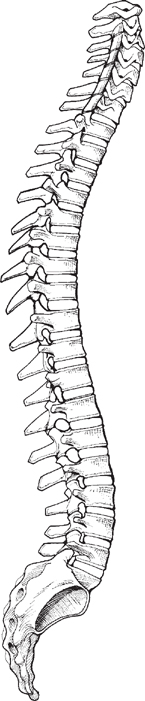
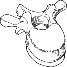
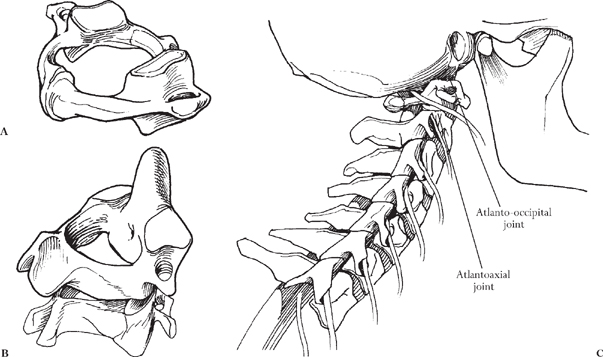
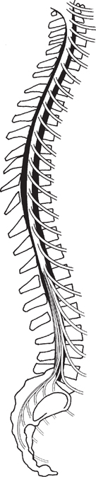
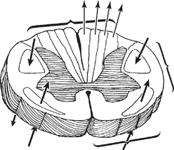
Chapter 1 CONTENTS II. BASIC NEUROLOGY OF THE SPINE VI. CLASSIFICATIONS OF MUSCLE GRADING VIII. REFLEX GRADING CLASSIFICATION SYSTEM Accurate diagnosis of spinal disease requires a thorough history, physical examination, and analysis of appropriate imaging studies, when appropriate. Often, the most important priority is to rule out alternative disease processes that can mimic spinal disorders. To accomplish these goals, a comprehensive understanding of basic spinal/neural anatomy is necessary as a first step. The vertebral column consists of 33 vertebrae, divided into 5 segments: cervical, thoracic, lumbar, sacral, and coccygeal (Fig. 1-1). There are seven cervical, 12 thoracic, 5 lumbar, 5 sacral, and 4 coccygeal vertebrae. The sacral and coccygeal vertebrae are usually fused to form the sacrum and the coccyx, respectively. The vertebrae of each segment are similar with several variations. The typical vertebra consists of a body, spinous process, two transverse processes, two pedicles, two arches, and two laminae (Fig. 1-2). Figure 1-1 Normal sagittal alignment and bony architecture of the cervical thoracic, lumbar, and sacrococcygeal spine. Figure 1-2 Normal vertebrae with body, processes, pedicles, and laminae. The two major exceptions are C1 and C2 (Fig. 1-3A, B). The first cervical vertebra, C1, is called the atlas and lacks the vertebral body. The atlas forms the atlanto-occipital joint with the occiput of the skull and contributes to flexion and extension of the neck (Fig. 1-3C). The second cervical vertebra is called the axis. Located on the superior side of the body of C2 is a bony protrusion called the dens or odontoid process. The dens fits into the ring of the atlas. Together, the atlas and axis make up the atlantoaxial joint, the major contributor toward cervical rotation. The vertebral bodies between C2 and S1 are each separated by a fibrocartilaginous intervertebral disk that acts as a buffer to mechanical shocks. Figure 1-3 (A) C1 and (B) C2 vertebrae. Note the difference in these two vertebrae, with the C1 being a ring and the C2 forming a bony peg that articulates with the ring of C1. (C) The atlantoaxial and atlanto-occipital joint contributes significantly to rotation and flexion/extension of the skull on the neck. The spinal cord resides within the vertebral foramen and extends from C1 to its ending, the conus medullaris, between L1 and L2 (Fig. 1-4). The filum terminale extends from the conus medullaris and attaches to the coccyx. The spinal cord is segmented, and 1 of the 31 pairs of spinal nerves exits from the spinal cord from each segment. There are 8 cervical, 12 thoracic, 5 lumbar, and 5 sacral pairs, and 1 coccygeal pair of spinal nerves. The spinal nerves of the cervical, thoracic, and lumbar cord exit through the intervertebral foramina; the sacral spinal nerves make up the cauda equina and exit through the sacral foramina. The spinal cord consists of a cellular core called gray matter surrounded by a fibrous layer called white matter. Spinal neurons, known as lower motor neurons, and interneurons reside in the gray matter. The axons of lower motor neurons together with afferent sensory neuron axons make up the white matter. The white matter is divided into four funiculi: right lateral, left lateral, ventral, and dorsal. Within the white matter are tracts of ascending and descending axons segregated into pathways of function (Fig. 1-5). Some of the most useful diagnostic pathways are the lateral spinothalamic tract, the dorsal columns, and the lateral corticospinal tracts. Figure 1-4 The neural anatomy is such that the spinal cord resides within the bony canal between C1 and L1 and/or L2. The filum terminale extends from the conus and attaches to the coccyx.
THE FUNDAMENTALS
Chapter 1
THE FUNDAMENTALS
BASIC ANATOMY OF THE SPINE
BASIC NEUROLOGY OF THE SPINE
Related posts:
 4 Physical Examination of the Lumbosacral Spine
 3 Physical Examination of the Thoracic Spine
 2 Physical Examination of the Cervical Spine
 Mesocircuit Mechanisms Underlying Recovery of Consciousness Following Severe Brain Injuries: Model and Predictions
 Working with the Microscope
Stay updated, free articles. Join our Telegram channel

Full access? Get Clinical Tree


