101 Median Nerve Entrapment at the Wrist

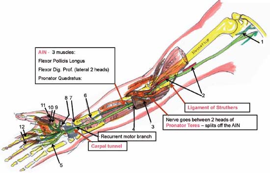
Case 101 Median Nerve Entrapment at the Wrist


Gaetan Moise and Christopher J. Winfree


Image Clinical Presentation


  • An 83-year-old right-handed woman without significant medical history presents with 3–4 weeks history of “tingling” in her first through third digits of the left hand.
  • Episodes are transient, mildly painful, and typically occur at night awakening her from sleep.

Image Questions


  1. What is the differential diagnosis?
  2. What are possible locations of median nerve entrapment?
  3. What is the most likely diagnosis?
  4. What special physical exam findings and maneuvers would support this diagnosis?
  5. What diagnostic tests should be ordered?
  6. What nonsurgical measures can be attempted to ameliorate the patient’s symptoms?
  7. When should surgery be considered?
  8. What are the surgical options?
  9. What complications are associated with carpal tunnel release?


Image

Fig. 101.1 Artist’s rendering of median nerve anatomical course and locations of entrapment. Sites of en trapment are highlighted in pink. AIN, anterior interosseous nerve; FDP or Flexor Dig. Prof., flexor digitorum profundus; PQ, pronator quadrates; AP, adductor pollicis; PT, pronator teres; F. Pol. Longus, flexor pollicis longus; FDS, flexor digitorum superficialis. (1) Lateral and medial cords join to form the median nerve. (2) Median nerve descends along the medial edge of the brachial artery. (3) Median nerve passes under the flexor digitorum superficialis (arcade forms the Sublimis Bridge). (4) Median nerve descends between flexor digitorum superficialis and profundus. (5) Sensory branches of the median nerve. (6) Anterior interosseous nerve (AIN) reaches deep to the pronator quadrates. (7) AIN gives off the wrist articular branches. (8) Recurrent motor branch variation. (9) Abductor pollicis. (10) Flexor pollicis. (11) Opponens pollicis. (12) Lumbricals I and II.


Image Answers


  1. What is the differential diagnosis?

    • Carpal tunnel syndrome (CTS)
    • Cervical radiculopathy
    • Brachial plexopathy
    • Supracondylar process syndrome (Struthers’ ligament)

  2. What are possible locations of median nerve entrapment?

    • Figure 101.1 shows the anatomic localizations of median nerve compression sites.

  3. What is the most likely diagnosis?

    • CTS results from median nerve compression at the carpal tunnel, a structure formed by several bones of the wrist and the transverse carpal ligament.1
    • The pathophysiology is thought to involve mechanical compression upon and ischemic changes within the median nerve.
    • Causes include repetitive hand movements, pregnancy, diabetes mellitus, acromegaly, hypothyroidism, multiple myeloma, amyloidosis, mucopolysaccharidosis, and rheumatoid arthritis.
    • It is the most common peripheral nerve compression syndrome and affects about 1–3% of adults.
    • Classic symptoms are restricted to a median nerve distribution distal to the carpal tunnel, but patients can present with proximal arm pain as well.

  4. What special physical exam findings/maneuvers would support this diagnosis?

    • Phalen’s test: patient rests elbows on exam table with forearms upright and allows wrist flexion with gravity assistance. Considered positive if median nerve distribution paresthesias commence or increase within 60 seconds.
    • Tinel sign: percussion of the median nerve at the wrist. Considered positive if reproduces paresthesias in a median nerve distribution.
    • Durkan’s compression test: application of continuous pressure to the median nerve at the transverse carpal ligament produces median nerve paresthesias, which are relieved by release of pressure.
    • Sensory: for example, two-point discrimination, monofilament testing, vibration detection

  5. What diagnostic tests should be ordered?

    • Electromyography and nerve conduction studies (EMG/NCS) are the standard diagnostic tests for CTS; conduction slowing of the median nerve across the wrist and denervational changes in the distal median-innervated muscles are commonly seen in CTS.
    • Cervical spinal imaging studies are unnecessary to make the diagnosis of CTS but are helpful to exclude other problems in the differential diagnosis such as cervical radiculopathy.
    • In this case, physical exam and NCS were consistent with the diagnosis of left-sided CTS; magnetic resonance imaging of the cervical spine was unremarkable.

  6. What nonsurgical measures can be attempted to ameliorate the patient’s symptoms?

    • Improve ergonomics associated with precipitating activities2
    • Avoid repetitive, stressful movements
    • Wrist splinting
    • Oral corticosteroids
    • Local injection of corticosteroids into carpal tunnel3
    • Nonsteroidal antiinflammatory medications, diuretics, vitamin supplements, and chiropractic manipulation are not thought to be helpful.

  7. When should surgery be considered?

    • Persistence of symptoms that do not improve with a month or so conservative management.3
    • Evidence of conduction slowing along the median nerve at the wrist on NCS.
    • Weakness of median-innervated muscles need not be present, but prompt earlier surgical intervention if present.
    • Denervational changes on needle EMG of median-innervated muscles distal to the carpal tunnel need not be present, but prompt earlier surgical intervention if present.

  8. What are the surgical options?

    • Mini-open carpal tunnel release involves decompressing the median nerve at the wrist through a 2-cm incision; although the patients may gently use the operated hand almost immediately, the incision requires a month or so of healing prior to aggressive work or rehabilitation activities (Fig. 101.2 and Fig. 101.3).4
    • Endoscopic carpal tunnel release involves decompressing the median nerve at the wrist through two 5-mm incisions; recovery times prior to return to work or aggressive physiotherapy are shorter after the endoscopic release compared with open release.4
    • Endoscopic carpal tunnel release is somewhat more expensive and has a higher risk of complications when performed by the inexperienced surgeon than open release.4

  9. What complications are associated with carpal tunnel release?

    • Injury to the main trunk median nerve, the palmar cutaneous branch, or the recurrent motor branch5
    • Incomplete release
    • Painful scar
    • Injury to the superficial palmar arch
    • Infection
    • Complex regional pain syndrome
< div class='tao-gold-member'>

Only gold members can continue reading. Log In or Register to continue

Stay updated, free articles. Join our Telegram channel

Jul 16, 2016 | Posted by in NEUROSURGERY | Comments Off on 101 Median Nerve Entrapment at the Wrist

Full access? Get Clinical Tree

Get Clinical Tree app for offline access