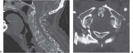
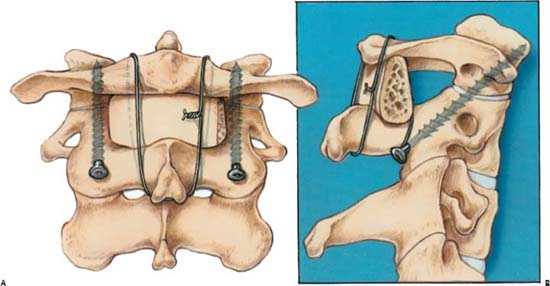
82 Atlantoaxial Instability

Case 82 Atlantoaxial Instability


Eric P. Roger and Edward Benzel


Image

Fig. 82.1 (A) Sagittal computed tomography (CT) reconstruction with myelographic subarachnoid contrast injection. (B) Axial CT nonmyelogram cut at the level of C1 through the dens.


Image

Fig. 82.2 Artist’s rendering of ideal trajectory of C1–C2 transarticular screws with (A) anteroposterior and (B) lateral illustrations. Posterior wiring with interspinous iliac graft is also depicted. Note that the posterior wiring of C1 to C2 may require a structurally intact arch of C1 and may not be feasible in some cases of Jefferson fracture (such as possibly in this case). (From Wolfla CE, Resnick DK. Neurosurgical Operative Atlas. Spine and Peripheral Nerves. New York: Thieme/American Association of Neurological Surgeons; 2006. Reprinted with permission.)


Image Clinical Presentation


  • An 83-year-old man presents with neck pain.
  • He had sustained a fall 6 months prior. There was no documentation of fracture at the time. He has been experiencing neck pain since the fall.
  • He denies any bladder or bowel dysfunction, gait or balance disorders.
  • He is admitted for generalized weakness and shortness of breath.
  • Upon further workup, he is found to be severely hyperkalemic with mental changes.
  • His past medical history is remarkable for placement of a cardiac pacemaker.

Image Questions


  1. What would be the sequence of radiologic investigations recommended for this patient?
  2. Interpret the images in Fig. 82.1.
  3. Is this an acute or chronic process? Why?
  4. How commonly are the pathologies at C1 and C2 associated?
  5. What are your therapeutic options at this point? How likely are they to be successful in achieving stability?
  6. Is spinal cord decompression indicated?
  7. What techniques are available for C1–C2 fixation? What factors would influence the decision making?
  8. Would an odontoid screw be a good surgical option in this patient?
  9. List the disadvantages of posterior C1–C2 fusion.
  10. What would you do if you had pulsatile bright red blood while drilling the trajectory for the placement of the transarticular C2 screw?

Image Answers


  1. What would be the sequence of radiologic investigations recommended for this patient?

  2. Interpret the images in Fig. 82.1.

    • Type II odontoid fracture with minimal translation, but ~60 degrees of posterior angulation
    • Soft tissue pannus behind the odontoid, partially indenting the cerebrospinal fluid (CSF) space, but not in direct contact with the cord (at least in this static CT image in supine position)
    • Axial image demonstrates anterior atlantal ring fracture consistent with a Jefferson fracture (posterior ring fracture not illustrated). This finding is suggested on Fig. 82.1A by the absence of the anterior ring on this midline cut.
    • Various levels of degenerative changes are noted through the rest of the cervical spine, without evidence of cord compression.

  3. Is this an acute or chronic process? Why?

    • These fractures are likely to be chronic because of the following:

      • The fracture line at the base of the dens appears sclerotic.
      • The fracture of the anterior atlantal arch is completely corticated.
      • Soft tissue pannus formation behind the odontoid is suggestive of chronic instability.
      • The patient presents with a fall 6 months prior, with neck pain since. There is no clinical evidence of an acute fall (i.e., don’t forget the clues given on history!).

  4. How commonly are the pathologies at C1 and C2 associated?

    • Axis fractures are relatively commonly associated with C1 fractures. Odontoid fractures (type II or III) are associated to C1 fractures in up to 53% of cases, and up to 26% with hangman’s fractures. They are reported to have a higher morbidity and mortality rate. Treatment is primarily based on the character istics of the C2 fracture.2

  5. What are your therapeutic options at this point? How likely are they to be successful in achieving stability?

    • Conservative management:

      • Soft collar
      • Semirigid collar (Philadelphia, Aspen, Miami J, etc.)
      • Rigid cervicothoracic orthosis

        • Minerva brace
        • Halo

    • Surgical stabilization3

      • Occipitocervical fusion
      • Atlantoaxial fusion
      • Odontoid screw is not an option in chronic fractures.

    • As these fractures are chronic, they are highly unlikely to heal with any type of external immobilization. Stability can be obtained only by open fixation and bone grafting. Conservative management should be considered only for patients medically unfit for surgery or, obviously, for patients refusing surgery.
    • Halo bracing is poorly tolerated by geriatric patients. Furthermore, although solid fixation of the head and chest may be achieved, paradoxical “snaking” motion of the cervical spine may occur.
    • Open fixation with bone grafting has a high fusion rate (>90%), except in smokers or in patients on steroids.

  6. Is spinal cord decompression indicated?

    • No. As illustrated by the CT myelogram, although there is soft tissue pannus formation with indentation onto the CSF space, there is no active cord contact or compression.
    • Furthermore, the patient does not present with any signs of myelopathy.

  7. What techniques are available for C1–C2 fixation? What factors would influence the decision making?

    • Available techniques include the following3,4:

      • Spinous process wiring (requires a structurally intact arch of C1):

        • Gallie fusion
        • Brookes fusion
        • Modified Sonntag fusion

      • C1–2 fixation:

        • C1 and C2 pars screws
        • C1 pars and C2 pedicle screws
        • C1 pars and C2 translaminar/interlaminar screws
        • C1–C2 transarticular screws
        • C1–C2 Halifax clamps

      • Odontoid screw placement
      • Occiput to C2 fixation

    • C1–C2 spinous process wiring (Brooks, Gallie, Sonntag) is a good adjunct to transarticular screws or to a segmented fusion but is not biomechanically a very strong construct when used as standalone.5
    • Spinous process wiring may be performed in a variety of ways but requires the integrity of the lamina and spinous process of C2, as well as integrity of the posterior arch of C1. In this case, although no decompression is required, there is loss of integrity of the arch of C1 due to the Jefferson fracture.
    • C1–C2 transarticular screw placement and C1–C2 segmental fusion (C1 lateral mass screws, C2 pars interarticularis screws/C2 pedicle screw fixation) in conjunction with spinous process wiring results in biomechanically strong constructs with higher rates of successful fusion when compared with spinous process wiring alone.6
    • C1–C2 posterior fixation techniques do not rely on the integrity of the posterior elements. Nonetheless, they are not without risk. Specifically, the trajectory of the vertebral artery and bony anatomy of the C2 pars must be carefully studied on preoperative imaging. High-riding vertebral arteries or a thin C2 pars may preclude the placement of a transarticular screw (Fig. 82.2 for ideal screw trajectory). If the arches of C1 or C2 are compromised (making spinous process wiring procedure not feasible), an onlay fusion laterally at the level of the articulating processes may alternatively be used in conjunction with C1–C2 transarticular screw placement.
    • C2 fixation using translaminar/interlaminar screws significantly reduces the risks to the vertebral artery, although epidural cortical breach is possible.
    • Preservation of C1–C2 rotation can also be achieved with a halo; however, this comes with associated morbidity in the elderly population.7

  8. Would an odontoid screw be a good surgical option in this patient?

    • Odontoid screws are indicated for acute type II odontoid fractures only. The screw is intended to “pull” the free dens against the C2 body, encouraging healing by compression along the fracture line. It is not a good option in this case.

  9. List the disadvantages of posterior C1–C2 fusion.

    • Disadvantages of C1–C2 posterior fixation include the loss of 50% of head rotation.
    • Furthermore, there is the potential for vertebral artery injury as well as significant venous plexus bleeding during lateral dissection of the C1–C2 joint.
    • The vertebral artery may be located in the path of a transarticular screw on preoperative images on at least one side in 18–23% of patients.8
    • Rate of intraoperative vertebral artery injury with this approach is estimated to be 1.7–4.1%.9
    • When C1–C2 spinous process wiring is utilized as a stand-alone technique, there is a high rate of pseudoarthrosis approaching 30% without halo fixation.9

  10. What would you do if you had pulsatile bright red blood while drilling the trajectory for the placement of the transarticular C2 screw?

    • This is consistent with damage to the vertebral artery. The only way to stop the bleeding is placement of the screw. If this is the first screw (and you still need to do the other side) you should not place the second screw for fear of causing bilateral vertebral artery damage potentially leading to devastating neurologic sequelae including death. Unilateral fixation is then accepted. Spinous process wiring may be considered.9
    • If such bleeding occurs while working on the second screw, then placement of the screw is again the only way to stop the bleeding.
    • Postoperative vertebral angiogram is indicated if damage to the vertebral artery is suspected. Vertebral artery stenting may be considered.
< div class='tao-gold-member'>

Only gold members can continue reading. Log In or Register to continue

Stay updated, free articles. Join our Telegram channel

Jul 16, 2016 | Posted by in NEUROSURGERY | Comments Off on 82 Atlantoaxial Instability

Full access? Get Clinical Tree

Get Clinical Tree app for offline access