104 Right Axillary Mass with Tinel Sign

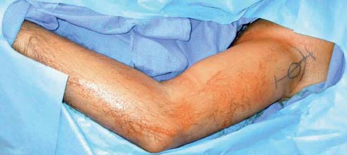
Case 104 Right Axillary Mass with Tinel Sign


Deepa Danan and Christopher J. Winfree


Image

Fig. 104.1 Intraoperative photograph demonstrating location of the mass in the axilla of the right upper extremity (circle) and placement of the overlying incision. The arm is draped into the field to facilitate interpretation of intraoperative nerve stimulation. Specifically, the incision along the course of the nerve to permit tumor removal is located along a portion of the nerve that, when stimulated, does not produce muscle contractions, so that the risk of neurologic injury is reduced.


Image Clinical Presentation


Image Questions


  1. What is in this patient’s differential diagnosis?
  2. What components of the history and physical examination suggest a benign versus malignant diagnosis?
  3. Are you going to order any tests? If so, which one(s)?
  4. What is the appropriate course of treatment?
  5. What is the typical outcome for benign lesions in this location?
  6. What is the typical outcome for malignant lesions in this location?

ImageAnswers


  1. What is in this patient’s differential diagnosis?

  2. What components of the history and physical examination suggest a benign versus malignant diagnosis?

  3. Are you going to order any tests? If so, which one(s)?

    • Electromyography and nerve conduction studies (EMG/NCS) may help localize the site of origin of the tumor, and document any neurologic injury caused by the tumor, but are not crucial in the workup of these patients.
    • Imaging studies are more important than EMG/NCS; magnetic resonance imaging (MRI) using T1-weighted, fat-saturated, gadolinium-enhanced images generally show peripheral nerve tumors in this location quite nicely.

  4. What is the appropriate course of treatment?

    • Brachial plexus exploration for gross total excision of the lesion is performed to ascertain a diagnosis, establish prognosis, provide symptomatic relief, and prevent progression of disease.1
    • Benign lesions are generally easily removed from the parent nerve using microdissection techniques.
    • After appropriate identification and neurolysis of the involved neural element, a longitudinal incision over the length of the tumor is made; benign tumors are then easily separated from the nerve fascicles, and the attached nerve fascicles are divided (Figs. 104.2, 104.3, and 104.4).1
    • When dissection planes are difficult to obtain, or if the tumor is unusually adherent or grossly invading surrounding tissue planes, then malignancy is suspected and intraoperative frozen section is performed.
    • If malignancy is confirmed on biopsy, then attempts at resection are abandoned; once final pathology is confirmed in subsequent days, then adjuvant therapy, such as radiation, and/or chemotherapy may be indicated.2
    • When possible, limb-sparing oncologic resection is attempted; when limb-sparing techniques are not possible, the limb amputation is performed.3

  5. What is the typical outcome for benign lesions in this location?

    • Benign schwannomas are associated with about a 7% risk of neurologic injury at the time of excision, and have a very low recurrence rate.
    • Benign neurofibromas have a higher risk of neurologic injury (20%) and may have a somewhat higher recurrence rate.4

  6. What is the typical outcome for malignant lesions in this location?

    • Unfortunately, despite aggressive surgical and adjuvant therapies, malignant peripheral nerve sheath tumors remain quite lethal.
    • In one series, the recurrence rate was ~50% by 5 years.3
    • In another series, the mean survival was just over 2 years.1
< div class='tao-gold-member'>

Only gold members can continue reading. Log In or Register to continue

Stay updated, free articles. Join our Telegram channel

Jul 16, 2016 | Posted by in NEUROSURGERY | Comments Off on 104 Right Axillary Mass with Tinel Sign

Full access? Get Clinical Tree

Get Clinical Tree app for offline access