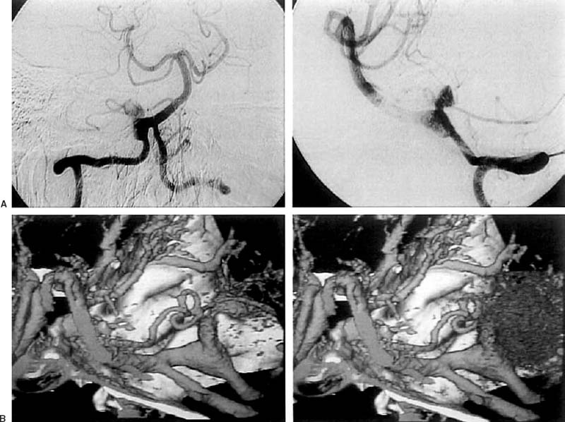
19 Diagnosis Partially thrombosed giant dissecting aneurysm of the vertebral artery Problems and Tactics Keywords Aneurysms, vertebral artery, arterial reconstruction, clipping A 56-year-old woman was admitted to a local hospital with history of sudden onset, headache and vomiting. She had developed giddiness a few days prior to this episode. Computed tomographic (CT) scan of the head done at the local hospital showed subarachnoid hemorrhage predominantly involving the right cerebellopontine angle. She was conscious but lethargic at the time of admission to our center. The Glasgow Coma Scale was 14 (3,5,6). Digital substraction angiography (DSA) demonstrated a dilated vertebral artery (VA) on the right side with a giant partially thrombosed aneurysm, which was confirmed by three-dimensional computed tomographic angiography (3D-CT) (Fig. 19–1). The patient was taken to surgery, which was performed in the left lateral position. The VA was exposed through the right lateral suboccipital craniotomy. The dilated vasa vasorums could be seen on the surface of the VA. The giant aneurysm of VA was gray in color suggestive of a dissecting aneurysm with an intramural thrombus. Temporary clipping of the proximal VA was done initially, followed by temporary clipping of the distal VA with the help of an endoscope (Fig. 19–2). The aneurysm was then opened and thrombectomy carried out. The thrombus was found to be attached to the aneurysm wall and was within the aneurysm. The aneurysmal cavity was examined using the endoscope, which revealed that the lumen of the VA was normal and only the dissecting portion contained the thrombus. The aneurysm and arterial reconstruction were clipped using a curved fenestrated clip and a curved nonfenestrated clip. Clip placement was checked using the endoscope and good blood flow was confirmed by intraoperative Doppler. The fenestrated clip were used to avoid inadequate closure of clipblades due to the presence of thickwall and thrombus. Postoperative recovery was uneventful and the patient was discharged without any neurological deficits. A postoperative angiogram showed good arterial reconstruction of the VA with mild dilation (Fig. 19–3). Giant aneurysms1–14 of the VA pose a formidable challenge to the vascular surgeon as well as to the endovascular surgeon. It is imperative that the attending surgeon considers all the treatment options. FIGURE 19–1 (A)
Partially Thrombosed Giant Dissecting Aneurysm of the Vertebral Artery—Treatment Strategies
Clinical Presentation
Discussion
![]()
Stay updated, free articles. Join our Telegram channel

Full access? Get Clinical Tree


