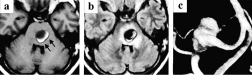
20 Diagnosis Thrombosed giant vertebral artery Problems and Tactics A case of a partially thrombosed giant aneurysm of the vertebral artery was presented. Despite no angiographic evidence of filling after endovascular treatment, the aneurysm continued to enlarge. Aneurysmectomy with removal of the coils resulted in a complete cure of symptoms. Intraoperative and histological findings suggested that neovascularization via vasa vasorum on the occluded vertebral artery provided a collateral supply to the unorganized thrombus in the aneurysm lumen, causing enlargement of the aneurysm. Keywords Thrombosed giant aneurysm, vertebral artery, coil embolization, vasa vasorum A 58-year-old female presented with a progressive swallowing disturbance, dysarthria, and truncal ataxia. T1-weighted magnetic resonance imaging (MRI) showed a giant thrombosed aneurysm compressing the brain stem, with a hyperintense area indicating the presence of a subacute clot within the aneurysm (Fig. 20–1A). On a fluid-attenuated inversion recovery (FLAIR) image, there was extensive edema in the brain stem (Fig. 20–1B). The right vertebral angiogram showed a small patent aneurysm lumen located immediately distal to the origin of the posterior inferior cerebellar artery (PICA) (Fig. 20–1C). The patient underwent endosaccular obliteration with occlusion of the parent artery and PICA using Guglielmi detachable coils (GDCs), resulting in complete angiographical obliteration of the aneurysm (Fig. 20–2A). Despite temporary relief of her symptoms and no evidence of recanalization on angiography, the aneurysm continued to enlarge with aggravation of surrounding brain stem edema (Fig. 20–2B) and recurrence of her symptoms, necessitating surgical treatment. The patient underwent a right suboccipital craniectomy and thrombectomy followed by partial aneurysmectomy. Of note, a markedly developed vasa vasorum was seen on the proximal vertebral artery (VA) and aneurysmal neck packed with coils (Fig. 20–3A). The aneurysmal wall near the neck also received an arterial supply from small branches of the right anterior inferior cerebellar artery (AICA) as well as the vasa vasorum from the parent artery (Fig. 20–3B). The vasa vasorum on the right VA seemed to derive from the dura close to its entrance to the intracranial cavity. The patent portion of the proximal VA as well as the origin of the PICA was occluded with a clip. When the middle of the adjacent VA segment, completely occluded on the angiogram, was cut with microscissors, there was oozing of blood through the coils packed within the parent artery on the aneurysm side. The VA immediately distal to the neck of the aneurysm was dissected and also occluded by clipping. When the aneurysm was opened, the packed coils were found to be embedded in the partially organized thrombus. The packed coils and friable clot were removed, leaving the outer shell of the aneurysm attached to the brain stem. FIGURE 20–1 A partially thrombosed giant vertebral artery (VA)–posterior inferior cerebellar artery (PICA) aneurysm (arrows). (A) T1-weighted magnetic resonance imaging (MRI) showing a thrombosed giant aneurysm at the right VA–PICA junction, compressing the brain stem. (B) Fluid attenuated inversion recovery (FLAIR) MRI showing marked edema in the brain stem surrounding the aneurysm. (C) The right VA three-dimensional digital angiogram showing a patent portion of the aneurysm, projecting superiorly, at the VA–PICA junction.
Collateralization via Vasa Vasorum: A Determinant of Therapeutic Efficacy of Coil Embolization of the Thrombosed Giant Aneurysm of the Vertebral Artery
Clinical Presentation
Surgical Technique
Histopathological Findings
Related posts:
 28 Dominant Hemisphere Insular Tumor
 9 Large Aneurysm of the Internal Carotid Artery Obliterated with Seven Fenestrated Clips under Intraoperative Monitoring of Anterior Choroidal Arterial Blood Flow Insufficiency
 8 Emergency Recanalization of the Inferior M2 Trunk by Arteriotomy and Retrieval of Migrated Guglielmi Detachable Coil, and Clipping of Superior Wall Aneurysm of Proximal Internal Carotid Artery
 41 Olfactory Neuroblastoma with Brain Invasion
 45 Clinoidal Meningioma Encasing the Internal Carotid Artery
 70 Fibrous Dysplasia of the Paranasal Sinuses and Anterior Cranial Base
Stay updated, free articles. Join our Telegram channel

Full access? Get Clinical Tree


