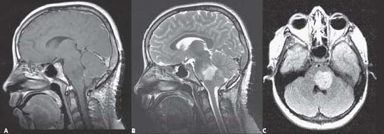
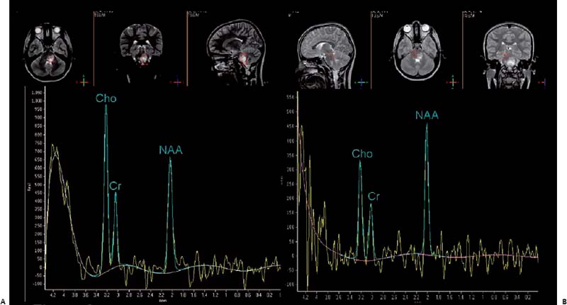
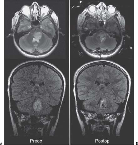
58 Brainstem Glioma 1: Pons

Case 58 Brainstem Glioma 1: Pons


Abdulrahman J. Sabbagh, Ayman Abdullah Albanyan, Mahmoud A. Al Yamany, Reem Bunyan, Ahmed T. Abdelmoity, and Lahbib B. Soualmi


Image

Fig. 58.1 (A) Brain sagittal T1-weighted magnetic resonance image (MRI) with gadolinium, (B) sagittal T2-weighted MRI and (C) axial fluid-attenuated inversion-recovery (FLAIR) image through the pons.


Image

Fig. 58.2 Magnetic resonance spectroscopy showing voxel configuration taken within the tumor (A) and within normal pons (B).


Image Clinical Presentation


Image Questions


  1. Describe the MRI.
  2. Describe the MR spectroscopy images. What is your differential diagnosis?
  3. How can you anatomically explain the 6th and 7th CN palsies?

    Over the following months her diplopia becomes worse and on examination, her 6th and 7th CN palsy becomes complete and now she has a significant pronator drift and mild swallowing difficulties. A repeat MRI study shows that the lesion is enlarging.


  4. If you chose to operate, what would be the aim of the surgery?
  5. How would you approach this lesion?
  6. What neurophysiologic modalities would you utilize during surgery?
  7. What are the safe entry zones into the floor of the 4th ventricle?

    You were able to resect close to 60% of the tumor (Fig. 58.3 shows the postoperative MRI study). The surgery is performed in the intraoperative MRI (iMRI) suite using intraoperative neurophysiology monitoring (IOM) (IOM electrodes were tested for MRI compatibility).


      Her swallowing ability returns to normal, but she has some balance issues that improve with physiotherapy. The 6th and 7th CN palsy remains. Pathologic tissue diagnosis comes back as grade II diffuse astrocytoma.


  8. How would you further manage this case?
  9. Classify pontine tumors.


Image

Fig. 58.3 Comparison bet ween (A) preoperative and (B) postoperative magnetic resonance images showing axial T2-weighted and coronal fluid-attenuated inversion-recovery images taken at the level of the pontine tumor.


Image Answers


  1. Describe the MRI.

  2. Describe the MR spectroscopy images. What is your differential diagnosis?

    • MR spectroscopy shows increased choline/creatine (Cho/Cr) and decreased N-acetyl aspartate/creatine (NAA/Cr) ratio within the lesion.
    • These findings are consistent with low-grade gliomas or demyelination processes.1,2

  3. How can you anatomically explain the 6th and 7th CN palsies?

    • This lesion involves the facial colliculus, which is formed by the facial motor fibers as they circle around the abducens nucleus in the dorsum of the pons.3
    • A lesion in the facial colliculus affects both the facial motor fibers and the abducens nucleus (Fig. 58.4).

  4. If you chose to operate, what would be the aim of the surgery?

    • The aim of surgery is 2-fold:

      • Decompression of the pons
      • Obtaining tissue for diagnosis

  5. How would you approach this lesion?

    • Suboccipital craniotomy–vermis-sparing telovelar approach
    • The infrafacial triangle may be utilized for approaching the tumor. As this lesion is occupying the facial colliculus and is pointing to the floor of the 4th ventricle, this lesion should be approached through the infrafacial triangle (that can be found by mapping or measurements) and/or the area closest to the surface of the 4th ventricle (Fig. 58.4).46
    • This approach would be further evaluated by use of neuronavigation and microscopy (Fig. 58.5).
    • In this particular case, an 8-mm area from the presumed midline and just below the striae medullares was used as the center of the infrafacial triangle and the closest part of the tumor to the 4th ventricle floor.

  6. What neurophysiologic modalities would you use during surgery?

    • Three modalities are available to monitor this patient7,8 (electrodes were checked and tested on a volunteer for MRI compatibility and the patient consented to monitoring):

      • Brainstem auditory evoked responses
      • Sensory evoked potentials
      • Motor evoked potentials
      • Fourth ventricular floor mapping

  7. What are the safe entry zones into the floor of the 4th ventricle?

    • Safe entry zones into the floor of the 4th ventricle include (Fig. 58.4)

      • Suprafacial (supraabducental) triangle: a triangle measuring around 16 mm in longest diameter. It is located above the facial colliculus and 5 mm from the midline (to avoid the medial longitudinal fascicle [MLF]). Its upper and narrower angle is below the trochlear nucleus.5,6
      • Infrafacial (infraabducental) triangle: a smaller triangle measuring less than 9 mm located just below the facial colliculus. It is narrow as it is located between the MLF medially and the facial nucleus laterally.5,6

  8. How would you further manage this case?

    • Management plan includes

      • For the residual tumor: conformal radiation or gamma knife treatment to the pons can be given postoperatively. This can be followed by serial MRI and close follow-ups.9,10
      • Note that, however, due to the relative risk and safety issues involved in using gamma knife treatments to the pons, some would not consider this option unless the lesion was exophytic from the pons.
      • For the facial palsy: teardrops and eye protection. One may resort to partial or gold-weight tarsorrhaphy in some cases to avoid corneal abrasions and ulcers. Facial-nerve reanimation procedures can also be tried.
      • For the gait disturbances: continued inpatient or outpatient rehabilitation

  9. Classify pontine tumors.

    • Classification of pontine tumors: Pontine tumors can be classified by type of tumor or growth pattern11:

      • Diffused pontine tumors: usually malignant and difficult to delineate from neighboring pontine parenchyma. Usually low or isointense signal on T1-weighted MRI and may have an increased signal on T2-weighted MRI. Hyperintensity on T1-weighted MRI is usually due to hemorrhagic change. Further enhancement in these diffuse tumors may be a sign of actual malignant degeneration.
      • Focal pontine tumors: well-demarcated and hypoor isointense signal on T1-weighted and high signal intensity on T2-weighted MRIs.
      • Exophytic pontine tumors: almost always dorsally exophytic into the 4 th ventricle and may be benign or malignant.12,13
< div class='tao-gold-member'>

Only gold members can continue reading. Log In or Register to continue

Stay updated, free articles. Join our Telegram channel

Jul 16, 2016 | Posted by in NEUROSURGERY | Comments Off on 58 Brainstem Glioma 1: Pons

Full access? Get Clinical Tree

Get Clinical Tree app for offline access