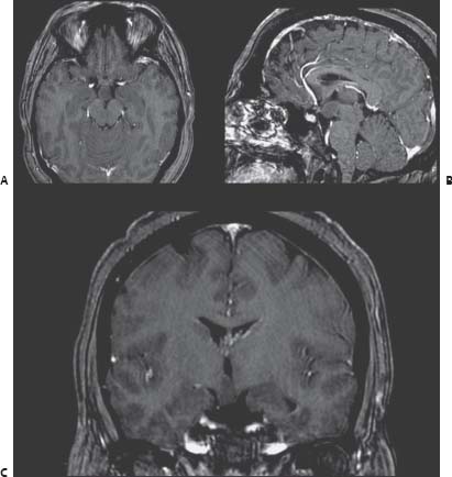
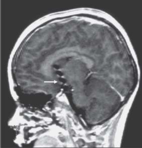
Case 65 Hypothalamic Hamartoma Fig. 65.1 T1-weighted magnetic resonance images of the brain with contrast, relevant (A) axial, (B) sagittal, and (C) coronal slices are shown. Fig. 65.2 T1-weighted sagittal magnetic resonance images of the brain showing depth electrode within the hypothalamic hamartoma.
 Clinical Presentation
 Questions
 Answers
< div class='tao-gold-member'>
65 Hypothalamic Hamartoma
Only gold members can continue reading. Log In or Register to continue

Full access? Get Clinical Tree


