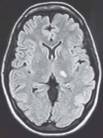
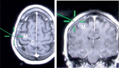
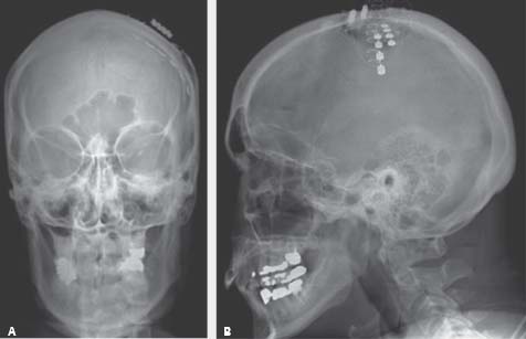
Case 77 Central Poststroke Pain Fig. 77.1 Magnetic resonance imaging fluid-attenuated inversion-recovery without contrast of the head demonstrating the small, left-sided thalamic infarct. Fig. 77.2 Intraoperative frameless stereotaxis magnetic resonance image demonstrating the localization of the central sulcus (C) and motor cortex (M). Identification of these landmarks facilitates the planning of a small craniotomy directly over the area of interest. Once the craniotomy is performed, somatosensory evoked potential phase reversal is performed to confirm the exact location of the central sulcus. The location of the facial and upper extremity motor cortex is confirmed when muscle contractions are elicited by transdural electrical stimulation. Once the array of stimulator electrodes is secured to the dura overlying the motor cortex, stimulation is increased until muscle contractions are noted, confirming the motor threshold. Postoperatively, stimulation intensities are kept well below the motor threshold to reduce the risk of postoperative seizures.
 Clinical Presentation
 Questions


![]()
Stay updated, free articles. Join our Telegram channel

Full access? Get Clinical Tree


