80 Neuronavigation and Intraoperative Imaging

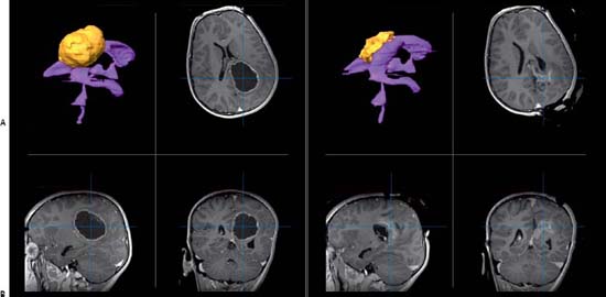
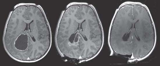
Case 80 Neuronavigation and Intraoperative Imaging


Lahbib B. Soualmi and Abdulrahman J. Sabbagh


Image

Fig. 80.1 (A) Magnetic resonance imaging (MRI) preoperative, three-dimensional (3D) reconstruction (top left) with ventricular system in purple and tumor in yellow. T1-weighted MRIs preoperative with contrast showing tumor in axial (top right), sagittal (bottom left), and coronal (bottom right) images. (B) MRI intraoperative, 3D reconstruction (top left) with ventricular system in purple and tumor in yellow. T1-weighted MRIs intraoperative with contrast showing tumor in axial (top right), sagittal (bottom left), and coronal (bottom right) images.


Image

Fig. 80.2 Intraoperative axial T1-weighted magnetic resonance images with contrast showing progression of tumor resection.


Image Clinical Presentation


Image Questions


  1. How will you plan the surgery?
  2. What are the characteristics of images needed for navigation in general?
  3. What are the types of information that can be utilized for navigation?
  4. How does neuronavigation work?
  5. What are the types of registration available?
  6. What are the existing types of localizers?
  7. How do you classify intraoperative imaging?
  8. Give examples where intraoperative imaging may be useful.
  9. Compare the advantages and disadvantages of intraoperative computed tomography (iCT) and intraoperative MRI (iMRI).

ImageAnswers


  1. How will you plan the surgery?

  2. What are the characteristics of images needed for navigation in general?

    • Three-dimensional (3D) global acquisition: Contiguous slices covering the whole region of interest such as the head or spine.

      • Anatomic isotropic scan with high resolution: Fine cuts are required to be able to create an accurate volumetric data.
      • After computerized fusion, the functional data can then be superimposed on the anatomic data.

  3. What are the types of information that can be utilized for navigation?

    • Anatomic information3,4

      • MRI

        • T1-weighted, ± contrast, T2-weighted, inverse recovery, fluid-attenuated inversion-recovery sequences, etc.
        • Diffusion tensor imaging, tractography
        • MR angiography, MR venography

      • CT

        • CT ± contrast,
        • CT angiography (CTA)

      • Ultrasound 3D acquisition

        • B-mode information
        • Doppler information

      • Conventional biplanar angiography

    • Functional information58

      • Positron emission tomography (PET)
      • Single photon emission computerized tomography
      • Functional MRI (fMRI)
      • Magnetoencephalography (MEG)
      • Cortical electroencephalography
      • Depth electrode recordings
      • Transcranial magnetic stimulation

    • Biochemical information

      • MR spectroscopy
      • Other chemical imaging modalities

  4. How does neuronavigation work?

    • The following are steps required for the workings of neuronavigation.912

      • Image acquisition (Fig. 80.3)
      • Image data analysis: anatomical ± functional data analysis—segmentation and 3D reconstruction and volume rendering of objects of interest
      • Fusion and superimposition of different anatomical ± functional images (Fig. 80.4): The example given is PET data superimposed on MRI data.
      • Registration: correlating and alignment of the 3D volumetric data with the actual patient (brain or spine)
      • Tracking: The neuronavigation platform should track the patient and surgical tools through the available localizer. This is done through different available tools (Fig. 80.5).

  5. What are the types of registration available?

  6. What are the existing types of localizers?

    • Optical localizers (using an infrared camera)14

      • Passive tracking: Using reflective spheres that are placed on the reference frame (patient), probes, and/or any surgical instrument (Fig. 80.5).
      • Active tracking: The reference frame (patient) and probes are equipped with light emitting diodes.

    • Magnetic localizers: Magnetic signals that are emitted by the probe and picked up by a receiver indicating its location in space. This modality avoids the disadvantage of a clear line of sight required for optical localizers.16
    • Mechanical localizers: Articulated arms equipped with transducers giving the angular position.
    • Ultrasound localizers: Navigation probes transmit ultrasonic signals that are picked up by a receiver indicating its location in space.

  7. How do you classify intraoperative imaging?

    • Real time3,17

      • Intraoperative ultrasound (iUS)
      • Intraoperative angiography
      • Fluoroscopy

    • Preacquired11

      • iMRI
      • iCT

  8. Give examples where intraoperative imaging may be useful.

    • Here are some examples of the use of intraoperative imaging.

      • iMRI, iCT, or iUS for detection or residual tumor18
      • iMRI, iCT, or iUS to compensate for brain shift
      • Reregister using intraoperative landmarks if the patient moves during surgery
      • iMRA, iCTA, or intraoperative angiography after clipping aneurysms or resecting arteriovenous malformations from the brain or spinal cord17,19
      • iUS to localize ventricles, cysts, and lesions in the brain and spinal cord
      • iUS for recalibration of navigation
      • iCT or iMRI for immediate localization of depth electrodes, strips, and grids (in epilepsy surgery), or deep brain stimulators (in functional and behavioral neurosurgery) to readjust them if needed20

  9. Compare the advantages and disadvantages of iCT with iMRI.

    • See Table 80.12024
< div class='tao-gold-member'>

Only gold members can continue reading. Log In or Register to continue

Stay updated, free articles. Join our Telegram channel

Jul 16, 2016 | Posted by in NEUROSURGERY | Comments Off on 80 Neuronavigation and Intraoperative Imaging

Full access? Get Clinical Tree

Get Clinical Tree app for offline access