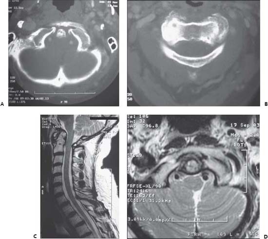
Case 84 Basilar Invagination Fig. 84.1 Cervical spine imaging with (A) axial computed tomography at the level of the foramen magnum and (B) at the atlantoaxial junction. (C) T2-weighted sagittal magnetic resonance image (MRI) and (D) axial MRI at the level of the foramen magnum.
 Clinical Presentation
 Questions
 Answers
< div class='tao-gold-member'>
Only gold members can continue reading. Log In or Register to continue


