87 Cervical Spondylotic Myelopathy

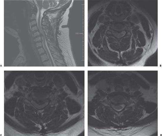
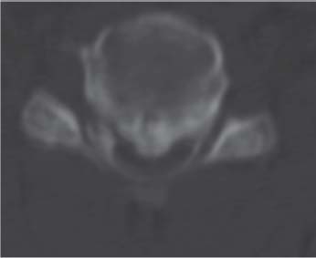
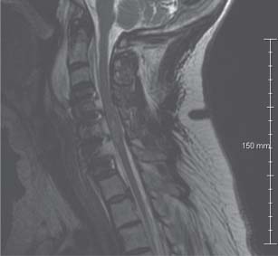
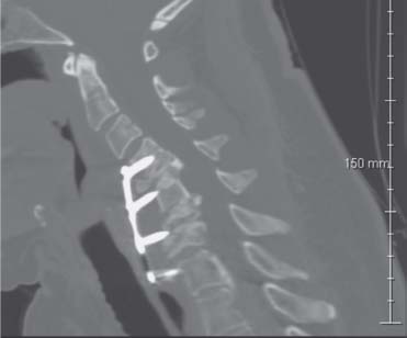
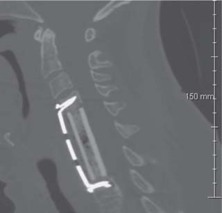
Case 87 Cervical Spondylotic Myelopathy


Remi Nader


Image Clinical Presentation


  • A 53-year-old woman presents with neck pain radiating to both shoulders as well as to the left arm and right hand.
  • She also complains of numbness and tingling in both upper extremities. Extending the neck slightly relieves some of the pressure; however, flexing the neck aggravates the pain.
  • There is an extensive medical history of hypertension, stroke, diabetes mellitus, asthma, and congestive heart failure.
  • Physical examination reveals some longstanding left-sided weakness (graded 4/5 on the Medical Research Council [MRC] motor scale) associated with her previous stroke (a few years ago).
  • Sensory examination reveals decreased sensation along the left side throughout, to temperature as well as de creased sensation bilaterally in a C5–C6 distribution to temperature. Reflexes are 1–2+ symmetric with no Hoffmann and no Babinski’s signs. Gait reveals a mild spasticity.
  • Magnetic resonance imaging (MRI) is ordered.


Image

Fig. 87.1 Magnetic resonance imaging of the cervical spine with T2-weighted images: (A) Midsagittal cut and axial cuts through the disk spaces at (B) C4–C5, (C) C5–C6, and (D) C6–C7.


Image Questions


  1. Interpret the MRI (Fig. 87.1).
  2. Give a differential diagnosis. What is the most likely diagnosis?
  3. What further studies would you like to obtain?
  4. A computed tomography (CT) scan of the cervical spine is obtained and shown in Fig. 87.2. Describe the pertinent finding.
  5. Briefly describe the pertinent points of your operative approach.
  6. Immediately postoperatively, she awakens with bilateral upper extremity profound weakness in triceps, grips, and interossei. Lower extremity strength is unchanged. Give a differential diagnosis. What is the most likely diagnosis?
  7. What is your management now?
  8. You immediately obtain an MRI of the cervical spine (Fig. 87.3); interpret it.
  9. How do you treat the patient given the MRI findings?

    She improves and is discharged home after a short rehabilitation stay. She then sustains a fall 6 weeks post-operatively and presents with worsening bilateral lower extremity weakness to the point where she is unable to ambulate. You obtain the following CT scan (Fig. 87.4).


  10. Interpret the CT scan (Fig. 87.4).
  11. What is your management now?
  12. Describe your treatment options and a surgical approach.

    After a long discussion of the different treatment options with the patient, you elect to perform a multilevel corpectomy and place the patient in a halo postoperatively. She does well and her strength is improved postoperatively. You obtain a CT scan, shown in Fig. 87.5.


Image

Fig. 87.2 Axial computed tomography scan at C5–C6 level.


Image

Fig. 87.3 Sagittal T2-weighted magnetic resonance image of cervical spine obtained immediately postoperatively.


Image

Fig. 87.4 Computed tomography scan sagittal reconstructed images through cer vical spine.


Image

Fig. 87.5 Computed tomography scan sagittal reconstructed images through cervical spine obtained after the second procedure, showing repair of the kyphosis, alignment restoration, and decompression of the spinal cord.


ImageAnswers


  1. Interpret the MRI (Fig. 87.1).

    • There is a large C5–C6 herniated nucleus pulposus with severe spinal cord compression at that level as well as a kyphotic deformity at the same level.
    • There is also a smaller C4–C5 and C6–C7 herniated nucleus pulposus with some cord compression at these two levels.
    • There is possibly an osteophytic spur or ossified posterior longitudinal ligament (OPLL) at C5–C6

  2. Give a differential diagnosis. What is the most likely diagnosis?

    • Most likely diagnosis is cervical spondylotic myelopathy.
    • Differential diagnosis based on clinical presentation includes the following (mnemonic is “CITTEN DIVA”)1:

      • Congenital: Chiari malformation, syringomyelia, narrow canal/short pedicles, mucopolysaccharidoses, kyphosis, os odontoideum
      • Infection: syphilis (infarction), postviral (herpes, varicella, cytomegalovirus), epidural empyema, vertebral osteomyelitis, acquired immunodeficiency syndrome- [AIDS-] related myelopathy, tuberculosis (Pott disease), parasitic cyst
      • Traumatic: spinal shock, epidural hematoma, basal skull trauma, electrical injury, bony fracture
      • Tumor: spinal cord tumor (extradural, intradural extramedullary, intramedullary), metastases, carcinomatous meningitis, paraneoplastic syndrome
      • Endocrine: Cushing disease, obesity (epidural lipomatosis), acromegaly, Paget disease
      • Nutrition/toxins: vitamin B12 deficiency, local anesthetics
      • Degenerative: spondylotic myelopathy, OPLL, disk herniation
      • Inflammatory: transverse myelitis, multiple sclerosis, Devic syndrome, Guillain-Barre syndrome, amyotrophic lateral sclerosis
      • Vascular: spinal epidural, subdural, or subarachnoid hemorrhage, stroke, spinal cord infarction (syphilis, aorta-clamping intraoperatively, hypotension, aortic dissection), arteriovenous malformation, radiation necrosis (causes microvascular occlusion), contrast infusion
      • Acquired: herniated disk, OPLL

  3. What further studies would you like to obtain?

    • Plain radiographs with flexion and extension views
    • CT scan of the cervical spine
    • Medical workup

      • Laboratory studies: complete blood count, electrolytes, coagulation profile, type and screen
      • Electrocardiogram
      • Chest radiograph
      • Cardiac echography
      • Pulmonary function tests

    • Consultation with Internal Medicine for surgical clearance

  4. A CT scan of the cervical spine is obtained and shown in Fig. 87.2. Describe the pertinent finding.

    • There is OPLL–segmental type at the level of C5–C6

  5. Briefly describe the pertinent points of your operative approach.

    • Anterior approach is described.

      • Anterior cervical diskectomy at C4–C5, C5–C6, C6–C7 may be performed in the standard Smith-Robinson technique.2
      • May need to do partial versus complete corpectomy of C5 and/or C6 for resection of OPLL3
      • Expect possible dural breach and anticipate its repair
      • Fusion with allograft and plate instrumentation
      • Use radiographic guidance and microscope
      • Use diamond burr on OPLL segment to avoid injuring spinal cord
      • May elect to use somatosensory and motor evoked potential monitoring intraoperatively
      • May elect to supplement with posterior C4–C7 lateral mass screws

    • Alternatively, a posterior approach may be used (however, this is not the author’s preferred approach, as the pathology is located anteriorly). This may include:

      • Cervical laminoplasty from C3–4 to C7, or
      • Cervical laminectomies from C4 to C7 ± lateral mass fusion

  6. Immediately postoperatively, she awakens with bilateral upper extremity profound weakness in triceps, grips, and interossei. Lower extremity strength is unchanged. Give a differential diagnosis. What is the most likely diagnosis?

    • Complications related to surgical technique4,5

      • Spinal cord edema with central cord syndrome (most likely)
      • Cord injury intraoperatively
      • Spinal epidural hematoma at surgical site
      • Graft dislodgment
      • Neck hematoma

    • Complications of anesthesia6

      • Spinal cord infarction due to generalized hypotension
      • Lingering effect of paralytics or anesthetics

    • Complications related to her medical conditions

  7. What is your management now?

    • Obtain an urgent MRI of the cervical spine.
    • Start the patient on steroids (dexamethasone 10 mg intravenously (i.v.) once then 6 mg i.v. every 6 hours or use the high-dose methylprednisolone [Solu-Medrol; Pfizer Pharmaceuticals, New York, NY] spinal cord injury protocol).
    • Admit to intensive care unit for monitoring.
    • Avoid hypotension (keep mean arterial pressure >85 mm Hg).
    • Call the operating room for possible return to explore the wound if a hematoma is suspected.

  8. You immediately obtain an MRI of the cervical spine (Fig. 87.3); interpret it.

    • MRI shows diffuse cord edema and thickening as well as a cord signal at the level of C5–C6.
    • Otherwise, no obvious hematoma is seen.
    • Good alignment and good placement of grafts are visualized.

  9. How do you treat the patient given the MRI findings?

    • Treat medically with steroids.4
    • There are no indications for surgical intervention.

  10. Interpret the CT scan (Fig. 87.4).

    • There is collapse of the construct with extrusion of the anterior instrumentation.
    • There are compression fractures of C5, C6, and C7 vertebral bodies
    • Kyphotic deformity is seen again at C5–C6.
    • Extrusion of the graft at C4–C5 and collapse of the graft at C6–C7 is observed.

  11. What is your management now?

    • The patient needs urgent surgical intervention (within 24 hours) for cord decompression and repair of the kyphotic deformity.
    • The extruded hardware also needs to be removed.
    • The compression fractures of the vertebral bodies need to be repaired and proper alignment needs to be restored.
    • Obtain further studies preoperatively.

      • Plain radiographs and MRI of the cervical spine
      • Preoperative medical clearance from Internal Medicine (urgent)

  12. Describe your treatment options and a surgical approach.

    • Options include5

      • Anterior decompression and internal fixation with grafting followed by posterior fixation (lateral mass screws and rods/plates) down to the upper thoracic area, and placement in a cervical collar. (Given the pathology, this remains the author’s preferred approach. However, this was not performed in this case due to patient-related factors and medical issues—these types of issues need to be documented in the patient’s chart very clearly.)
      • Anterior decompression and internal fixation with grafting followed by placement of a halo vest (or a sterna-occipital mandibular immobilizer cervicothoracic orthosis if the patient is unable to tolerate the halo, although less ideal)
      • Posterior decompression and/or fixation alone is not a good option as the pathology is mainly anterior and the kyphotic deformity is significant.
      • Placement of a collar or other soft brace is not an option after anterior fixation alone, as there is involvement of C7 vertebral body, which places the graft at a junctional level.

    • Anterior decompression and fixation would consist of7

      • Anterior corpectomy of C5, C6, and C7 vertebral bodies
      • Fibular strut graft placement or metallic cage placement
      • Use of bone autograft from vertebral bodies to help with arthrodesis
      • Anterior instrumentation from C4 to T1 with plate and screws
      • Intraoperative adjuncts are as described previously.
< div class='tao-gold-member'>

Only gold members can continue reading. Log In or Register to continue

Stay updated, free articles. Join our Telegram channel

Jul 16, 2016 | Posted by in NEUROSURGERY | Comments Off on 87 Cervical Spondylotic Myelopathy

Full access? Get Clinical Tree

Get Clinical Tree app for offline access