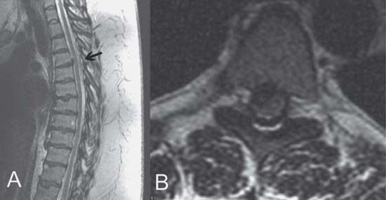
Case 92 Thoracic Disk Herniation Fig. 92.1 (A) Sagittal and (B) axial T2- weighted magnetic resonance images of the thoracic spine showing herniated disk (arrow) at the T4–T5 level.
 Clinical Presentation
 Questions
 Answers
< div class='tao-gold-member'>
92 Thoracic Disk Herniation
Only gold members can continue reading. Log In or Register to continue

Full access? Get Clinical Tree


