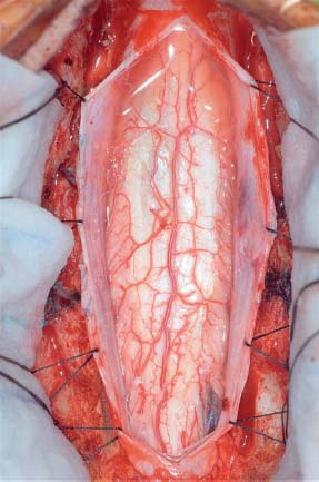
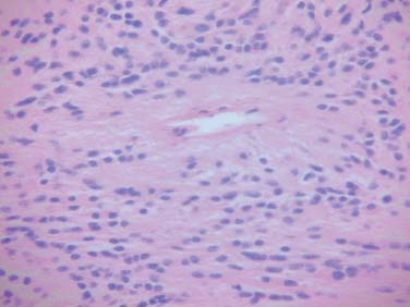
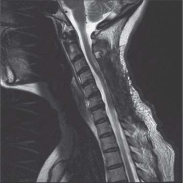
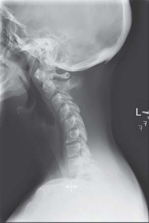
Case 94 Intramedullary Spinal Tumor Fig. 94.1 (A) Sagittal magnetic resonance imaging of the C-spine with T2-weighted image and (B) T1-weighted image with gadolinium revealing an intramedullary tumor. On T2-weighted images, the tumor is isointense to the spinal cord, with hypointensity (hemosiderin deposits) above and below the tumor and a hyperintense overlying syrinx. There is heterogeneous enhancement after gadolinium. Fig. 94.3 Histological section revealing perivascular pseudorosettes, characteristic of ependymoma. Fig. 94.4 Postoperative T2-weighted magnetic resonance imaging reveals no tumor mass. Fig. 94.5 Lateral C-spine radiograph demonstrating postlaminectomy kyphosis.

 Clinical Presentation
 Questions
 Answers
< div class='tao-gold-member'>
94 Intramedullary Spinal Tumor
Only gold members can continue reading. Log In or Register to continue

Full access? Get Clinical Tree


