96 Spinal Arteriovenous Malformation

Case 96 Spinal Arteriovenous Malformation


Bassem Sheikh


Image

Fig. 96.1 (A) T1-weighted and (B) myelographic magnetic resonance imaging, and (C) selective spinal digital subtraction angiography.
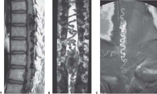

Image Clinical Presentation


  • A 32-year-old man presents with a 2-year history of progressive paraparesis.
  • He has weakness involving both lower limbs that is progressively increasing.
  • The patient also complains of back pain and bilateral leg numbness.
  • Disturbance of bladder function started 3 weeks prior to presentation.
  • He also shows loss of superficial sensation below the umbilicus. The vibration and position sensations are disturbed in both lower extremities.

Image Questions


  1. Provide a clinical explanation to the patient’s presentation.
  2. What is your differential diagnosis?
  3. Describe a classification of spinal arteriovenous malformations (AVMs).
  4. What is your diagnostic workup for spinal AVMs?
  5. Imaging studies are obtained and shown in Fig. 96.1. Describe the radiologic findings in these images.
  6. What are the therapeutic modalities that may be suggested for this patient?
  7. The patient asked you whether he would regain normal neurologic function after treatment. What is your answer to the patient?

Image Answers


  1. Provide a clinical explanation for the patient’s presentation.

    • This patient is suffering from a progressive myelopathy.
    • The myelopathy is most likely originating in the thoracic spine area given the clinical presentation.

  2. What is your differential diagnosis?

    • The differential diagnosis of spinal progressive myelopathy is broad and includes the following1,2:

      • Congenital conditions: Chiari malformation, syringomyelia, narrow canal/short pedicles, mucopoly-saccharidoses, kyphosis, os odontoideum
      • Infections: syphilis (infarction), postviral (herpes, varicella, cytomegalovirus), epidural empyema, vertebral osteomyelitis, acquired immunodeficiency syndrome (AIDS)-related myelopathy, tuberculosis (Pott disease), parasitic cyst
      • Traumatic conditions: spinal shock, epidural hematoma, electrical injury, bone fracture
      • Tumors: spinal cord tumor (extradural, intradural extramedullary, intramedullary), metastases, carcinomatous meningitis, paraneoplastic syndrome
      • Endocrine conditions: Cushing disease, obesity (epidural lipomatosis), acromegaly, Paget disease
      • Nutritional conditions or toxins: vitamin B12 deficiency, local anesthetics
      • Degenerative conditions: spondylotic myelopathy, ossified posterior longitudinal ligamen, disk herniation
      • Inflammatory or demyelinating conditions: transverse myelitis, multiple sclerosis, Devic syndrome, Guillain-Barre, amyotrophic lateral sclerosis
      • Vascular diseases: spinal epidural, subdural or subarachnoid hematoma, spinal cord infarction (syphilis, aorta clamping intraoperatively, hypotension, aortic dissection), AVM, radiation exposure, reaction to contrast infusion

  3. Describe a classification of spinal AVMs.

    • There have been several classification systems for spinal vascular malformation in the literature, however, they are broadly categorized into the following three sections3–6:
    • Dural arteriovenous fistula (DAVF): This is the most common type of malformation (Fig. 96.2).

      • It typically presents in older men.
      • It is usually found in the lumbar and thoracic spine.
      • This lesion consists of a small arteriovenous fistula within or just beneath the dura at the point where a feeding radicular artery enters the dura at the nerve root sleeve (at the level of the intervertebral foramen).
      • The venous outflow from the fistula, carrying arterialized blood, drains into the intradural venous plexus via the radiculomedullary vein along the dorsal surface of the spinal cord.
      • These low-flow fistulae produce venous hypertension (Foix-Alajouanine syndrome), which results in decreased spinal cord perfusion, resulting in intermittent and progressive neurologic deficits.

    • Perimedullary fistulas: located intradurally but in the extramedullary space

      • They usually present at the thoracolumbar region.
      • They are characterized by a single shunt without a nidus.
      • They occur between the spinal artery (anterior or posterolateral artery) and the spinal vein.
      • They may ascend rostrally forming craniocervical shunts, reaching even into the posterior cranial fossa.
      • They are located either on the ventral or dorsal surface of the spinal cord.

    • Intramedullary AVMs: These lesions are rare (Fig. 96.2).

      • They are supplied by the radiculomedullary artery or the spinal artery.
      • They are found partially or entirely within the substance of the spinal cord.
      • They are further subclassified according to their size and the location within the spinal cord.

        • Large complex metameric AVMs
        • Juvenile type
        • Combined intradural and extradural (intraspinal)

  4. What is your diagnostic workup for spinal AVMs?

    • Diagnostic evaluation includes the following:

      • Magnetic resonance imaging (MRI): Visualization of dilated spinal veins is possible, but is difficult in cases of DAVF because the only slightly dilated vessels and the fistula itself cannot be visualized. Increased intramedullary signal in T2-weighted images can reflect the edema due to chronic venous hypertension. Serpiginous areas of low signal owing to signal void in the draining vein may be demonstrated.
      • Magnetic resonance angiography
      • Myelography: has been replaced by the more informative MRI combined with MR myelographic effect. Computed tomography (CT) myelography can help define the feeding pedicle location.
      • Selective spinal angiography: This study remains the gold standard evaluation for spinal AVMs and is the only way of diagnosing and localizing the nidus in dural fistulae. It requires detailed catheterization of thoracic and lumbar arteries. If angiographic results are routinely negative, but there is a strong clinical suggestion of a fistula, it may be necessary to extend the investigation to the vertebral, the external carotid, or the sacral arteries.

  5. Imaging studies are obtained and shown in Fig. 96.1. Describe the radiologic findings in these images.

    • These images demonstrate the presence of an intradural, intramedullary AVM that is filling from a spinal artery and draining rostrally and caudally into draining veins.
    • Part of the nidus is present within the spinal cord substance.

  6. What are the therapeutic modalities that may be suggested for this patient?

    • Successful treatment in each individual spinal vascular malformation requires correct understanding of the lesion’s anatomic location and its angioarchitecture.
    • The therapeutic modality that should be suggested depends on the type of spinal vascular malformation.
    • The limitations of both surgery and endovascular embolization should be considered.
    • The following summarizes each treatment modality based on type of spinal AVM.3,7,8

      • DAVF

        • Surgical technique: surgical exposure to excise the fistula is a simple and successful treatment of spinal DAVFs.
        • Endovascular technique: As an alternative to surgical therapy, injection of N-butyl cyanoacrylate can be done after superselective catheterization of the radicular artery. This method has been reported to be successful and definitive in ~75% of cases. If the occlusion of the fistula is attained endovascularly with definitive embolization material, an operation is not necessary. Otherwise, removal of the partially embolized fistula has to be performed.

      • Perimedullary fistulas

        • Surgical elimination: This is an option for posteriorly located accessible small fistula.
        • Endovascular embolization: may be used in the case of ventrally situated fistulae that are inaccessible surgically and giant fistulae with multiple dilated feeders and draining veins

      • Intramedullary AVMs

        • Endovascular embolization: This is the modality of choice in the management of intramedullary AVMs. Most techniques use particulate embolization materials that result in reduction in the flow through the malformation, thus lowering the steal and ischemic effects on the spinal cord and the venous hypertension. However, particulate embolization is noncurative and revascularization of the malformation should be expected.
        • Surgical technique: Microsurgical therapy alone is sometimes technically difficult owing to the intramedullary and ventral location of the AVM. Figure 96.3 illustrates an intraoperative view of such an AVM.

  7. The patient asked you whether he would regain normal neurologic function after treatment. What is your answer to the patient?

    • Postoperative improvement of patients with neurologic deficits depends on preoperative duration of signs and symptoms and on the degree of disability.7,8
    • Improvements may usually involve both sensory and motor deficits.
    • The genitosphincteric disturbances have a much more severe prognosis and persist more often.
    • Because of progressive ischemic lesions of the spinal cord caused by chronic venous congestion, the shunt should be eliminated as early as possible.9
< div class='tao-gold-member'>

Only gold members can continue reading. Log In or Register to continue

Stay updated, free articles. Join our Telegram channel

Jul 16, 2016 | Posted by in NEUROSURGERY | Comments Off on 96 Spinal Arteriovenous Malformation

Full access? Get Clinical Tree

Get Clinical Tree app for offline access