2 Anatomy of the Nasal Cavity and Paranasal Sinuses
Carolina Martins, Luiz Felipe de Alencastro, Alberto Carlos Capel Cardoso, Alvaro Campero, Alexandre Yasuda, Jian Wang, Luiz Carlos de Alencastro, and Albert L. Rhoton, Jr.
 Introduction
The nasal cavity is the space between the nares and the choanae, separated along the midline by the nasal septum. It extends inferiorly from the palate up to the inferior surface of the cribriform plate, being on top of the oral cavity and anterior to the pharynx. The air space medial to the conchae and lateral to the septum is the common nasal meatus, which communicates at the lower edge of each concha with the superior, middle, and inferior meatus (Figs. 2.1, 2.2, 2.3, and 2.4).
From a surgical point of view, this space is a natural pathway to and from several adjacent compartments. The compartments that can be reached through the superior part of the nasal cavity are, from anterior to posterior, the frontal sinus cavity; the ethmoidal sinuses and the center of the anterior cranial base; and the sphenoid sinus cavity and sellar region. Laterally, the compartments related to the nasal cavity can be separated at the level of the middle meatus. Above this level, the nasal cavity can lead to the orbit and its contents; below, there is a two-way route to and from the maxillary sinus. At the level of the choanae, the lateral relationship of the nasal cavity includes also the pterygopalatine and infratemporal fossae.
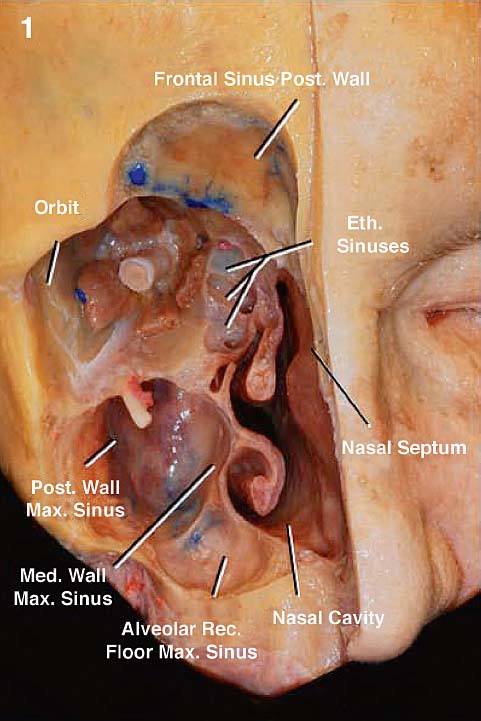
Fig. 2.1 The right side of the face has been dissected to expose the nasal cavity and its topographical relationships. Eth.: ethmoid, ethmoidal; Max.: maxillary; Med.: medial; Post.: posterior; Rec.: recess.
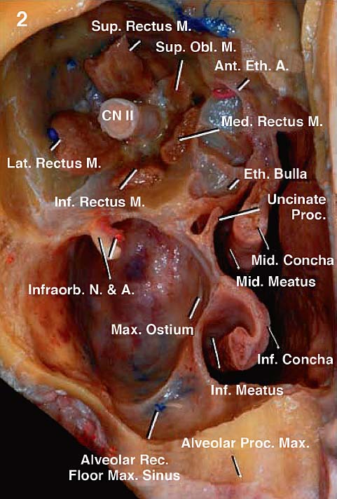
Fig. 2.2 Enlarged view of Fig. 2.1. The air space medial to the conchae and lateral to the septum is the common nasal meatus, which communicates at the lower edge of each concha with the superior, middle, and inferior meatus. The arrangement of the extraocular muscles, which attach posteriorly at the annulus of Zinn, helps divide the orbit into an intraconal and extraconal spaces. The optic nerve, the most important intraconal structure, is used as a reference to describe lesions located into this space. The maxillary sinus is a pyramidal cavity in the body of the maxilla. Its apex points laterally and extends into the zygomatic process. The base is the lateral wall of the nasal cavity below the middle meatus and presents the maxillary ostium. Its floor is the alveolar process of the maxilla. The alveolar canals transmit the posterior- superior alveolar nerves to the molar teeth and can protrude on the posterior wall of the sinus. The roof separates the sinus from the floor of orbit and presents the infraorbital canal for the infraorbital nerve and vessels. The anterior wall of the sinus forms the anterior surface of the body of the maxilla, faces the oral vestibule, and has been removed in this dissection. The uncinate process is a curved projection from the orbital plate of ethmoid at the medial wall of the maxillary sinus. A.: artery; Ant.: anterior; CN: cranial nerve; Eth.: ethmoid, ethmoidal; Inf.: inferior; Infraorb.: infraorbital; Lat.: lateral; M.: muscle; Max.: maxillary; Med.: medial; Mid.: middle; N.: nerve; Obl.: oblique; Proc.: process; Rec.: recess; Sup.: superior.

Fig. 2.3 The dissection has proceeded to the posterior limit of the nasal cavity. The orbit has been resected up to the apex, and the walls of the maxillary sinus, the nasal septum, and part of the palate have been removed. The pterygopalatine fossa, containing the pterygopalatine ganglion (*) and the infratemporal fossa, with structures related to the posterior wall of the maxillary sinus, have been exposed. The sphenoid sinus has been opened. The anterior and middle fossae have been exposed, and part of the tongue, the ramus, and the body of the mandible have been removed. At the level of the choanae, the lateral relationship of the nasal cavity includes the pterygopalatine and infratemporal fossae. Some of the adjacent compartments should be thought of as nasal cavity “amplifiers,” because, when working through them, several other areas can be reached. This is especially true for the maxillary and sphenoid sinus and the pharynx. The maxillary sinus expands the reach, particularly to the infratemporal and pterygopalatine fossae, as well as the orbit. The sphenoid sinus offers the possibility to reach the anterior fossa, the cavernous sinus, the petrous part of the temporal bone, and the middle and posterior fossae, besides the well-known path it provides to the sellae. The nasopharynx offers a path to the inferior clivus and the foramen magnum area, the craniovertebral junction, as well as the contents of the parapharyngeal and infrapetrosal spaces. A.: artery; Cav.: cavernous; CN: cranial nerve; Fiss.: fissure; Lat.: lateral; M.: muscle; Mand.: mandibular; Max.: maxillary; Med.: medial; Proc.: process; Pteryg.: pterygoid; Sphen.: sphenoidal.
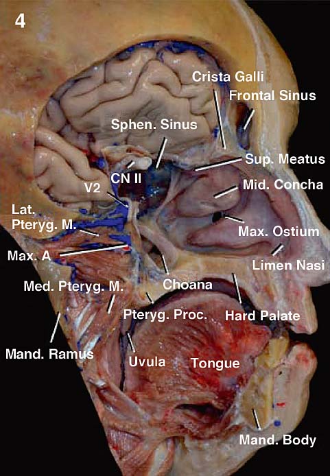
Fig. 2.4 The view along the lateral wall of the left nasal cavity. The nasal cavity proper extends from the limen nasi to the choana. The concha protrudes along this wall, delimiting the superior, middle, and inferior meatus. The frontal, anterior ethmoidal, and maxillary sinuses open into the middle meatus. A.: artery; CN: cranial nerve; Lat.: lateral; M.: muscle; Mand.: mandibular; Max.: maxillary; Med.: medial; Mid.: middle; Proc.: process; Pteryg.: pterygoid; Sphen.: sphenoidal; Sup.: superior; V2: second division trigeminal nerve.
Some of the adjacent compartments should be thought of as nasal cavity “amplifiers.” When working through these compartments, several other areas can be reached. This is especially true for the maxillary and sphenoid sinuses and the pharynx. The maxillary sinus expands the reach particularly to the infratemporal and pterygopalatine fossae, as well as the orbit. The sphenoid sinus offers the possibility of reaching the anterior fossa, cavernous sinus, petrous part of the temporal bone, and middle and posterior fossae, in addition to the well-known path it provides to the sellae. The nasopharynx offers a path to the inferior clivus and foramen magnum area, the craniovertebral junction, and the contents of the parapharyngeal and infrapetrosal spaces.
 Anatomy of the Nasal Cavity
The nasal cavity has a vestibule, which extends from the naris to the limen nasi and is related to the external nose. Posterior to the vestibule is the nasal cavity proper, extending from the limen nasi and piriform aperture to the choana (Fig. 2.4).
The piriform aperture is the anterior bony opening of the nasal cavity. It is formed by the nasal bones and frontal processes of the maxilla above and the alveolar processes of the maxilla below and laterally (Figs. 2.5 and 2.6). The anterior nasal spine is found at the inferior edge of the piriform aperture, at the meeting point of both maxillae. It relates superiorly to the free end of the septal cartilage, at the point that it unites with columella. The average superior width of the piriform aperture in adults is 16 mm; its breadth, 24 mm; and the height of the aperture, 29 mm.1 The cartilages of the external nose are arranged along the margins of the piriform opening and delimit the anterior openings of the nose—the nares.
The choana is the posterior limit of the nasal cavity and opens the nasal cavity proper to the nasopharynx (Figs. 2.3 and 2.4). Acting as a door, each choana is delimited superiorly by the sphenoid rostrum, superolaterally by the sphenoid process of the palatine bone, and laterally by the medial pterygoid plate and the perpendicular plate of the palatine bone. The inferolateral angle of the choana is smooth and formed by the meeting point between the perpendicular and horizontal plates of the palatine. Superomedially the choana is delimited by the ala of vomer and medially by the bony septum, formed by sphenoid rostrum and the crest united with the vomer. Inferomedially the choana is delimited by the nasal surface of the horizontal plate of the palatine bone at the level of the nasal crest (Figs. 2.7 and 2.8). The posterior nasal spine is the midline protrusion on the nasal surface of the horizontal plate of the palatine at the posteriormost point of the nasal crest. In adults, each choana has an average height of 25.5 mm and a width of 13.5 mm.1
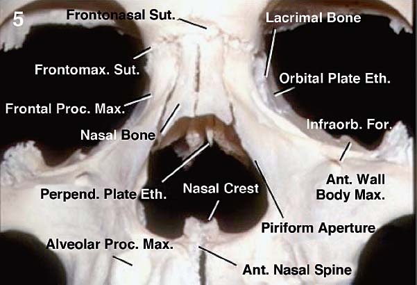
Fig. 2.5 The piriform aperture is the anterior bony opening of the nasal cavity. It is formed by the nasal bones and frontal processes of the maxilla above and the alveolar processes of the maxilla below and laterally. The nasal bones unite above with the frontal bone at the nasofrontal suture, and laterally with the frontal process of the maxilla at the nasomaxillary suture. The anterior nasal spine is found at the inferior edge of the piriform aperture, at the meeting point of both maxillae. Ant.: anterior; Eth.: ethmoid, ethmoidal; For.: foramen; Frontomax.: frontomaxillary; Infraorb.: infraorbital; Max.: maxillary; Perpend.: perpendicular; Proc.: process; Sut.: suture.
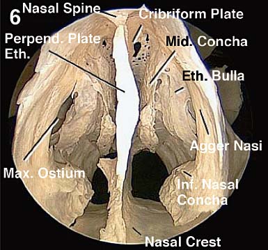
Fig. 2.6 Dry, articulated skull. Endoscopic view within the nasal cavity. The nasal spine of the frontal bone projects inferiorly and anteriorly. With the perpendicular plate of the ethmoid, the spine of the frontal bone supports the nasal bones, which lie upon them. Eth.: ethmoid, ethmoidal; Inf.: inferior; Max.: maxillary; Mid.: middle; Perpend.: perpendicular.
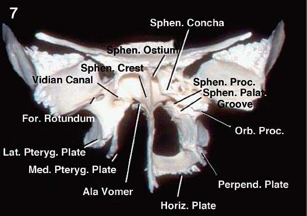
Fig. 2.7 The choanae are formed by the apposition of the sphenoid, vomer, and palatine bones. For.: foramen; Lat.: lateral; Horiz.: horizontal; Med.: medial; Orb.: orbital; Palat.: palatine; Perpend.: perpendicular; Proc.: process; Pteryg.: pterygoid; Sphen.: sphenoidal.
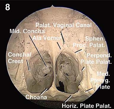
Fig. 2.8 A dry, articulated skull. Posterior endoscopic view of the choanae. Horiz.: horizontal; Med.: medial; Mid.: middle; Palat.: palatine; Perpend.: perpendicular; Proc.: process; Sphen.: sphenoidal.
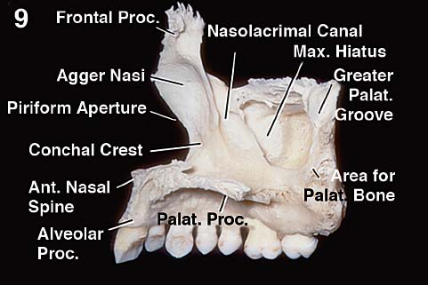
Fig. 2.9 The nasal surface of the maxillary body is responsible for most of the lateral wall of the nasal cavity. It presents the maxillary hiatus, a large opening on the posterosuperior part of the nasal surface of the maxillary body, which leads to the maxillary sinus. Inferior to the hiatus, the nasal surface contributes to the inferior meatus. Behind the hiatus, there is a rough surface for articulation with the perpendicular plate of the palatine bone. In this area, the greater palatine groove is converted into a canal by the articulation with the palatine. In front of the hiatus, a deep groove contributes to the formation of the nasolacrimal canal. The opening of the hiatus is reduced by the apposition of adjacent bones: the uncinate process of the ethmoid and descending part of the lacrimal above; the maxillary process of the inferior nasal concha below; and the perpendicular plate of palatine behind. The agger nasi, anterior to the hiatus is an elevation related to the anterior portion of the middle concha. Ant.: anterior; Max.: maxillary; Palat.: palatine; Proc.: process.
The lateral wall of the nasal cavity is formed by the nasal, maxilla, lacrimal, ethmoid, palatine, inferior nasal concha, and sphenoid bones (Figs. 2.9, 2.10, 2.11, 2.12, 2.13, 2.14, 2.15, 2.16, 2.17, 2.18, 2.19, and 2.20). The nasal bone contributes with the lateral part of its inferior surface to a small anterior area of the lateral wall. The maxilla provides the inner surface of the frontal process and the nasal surface of its body, to which the inferior nasal concha attaches. The lacrimal bone, through its medial or nasal surfaces, contributes to the formation of the nasolacrimal duct and attaches to the maxilla, ethmoid, and inferior nasal concha. The medial plates of the ethmoid bone contribute to the lateral walls of the upper part of the nasal cavity. Between the medial and lateral plates, also called the laminae papyracea or orbital plate, are the ethmoid labyrinths or lateral masses, formed by ethmoidal air cells. Two or three ethmoidal conchae may project into the cavity.
The palatine bone contributes to the lateral wall with the nasal surface of its perpendicular plate. The nasal surface has two horizontal ridges—the ethmoidal and conchal crests. The ethmoidal crest is the upper ridge and articulates to the middle concha of the ethmoid bone. The conchal crest is the most inferior ridge on the nasal surface of the perpendicular plate. Together with the conchal crest of the maxilla, it gives attachment to the inferior concha. Between the ethmoidal and conchal crests, the concave area forms part of the middle meatus. Similarly, below the conchal crest, a shallow depression forms the inferior meatus of the nasal cavity. The perpendicular plate attaches between the pterygoid plate of the sphenoid and the posteromedial margin of the nasal surface of maxilla. The upper end of the perpendicular plate has two processes separated by a groove. The anteriormost process is the orbital process of the palatine bone. It attaches to the maxilla, ethmoid, and sphenoid bones and encloses an air cell that may communicate with the posterior ethmoidal air cells or the sphenoid sinus. The posteriormost process, slightly deviated medially, is the sphenoidal process. By its posterosuperior surface, the sphenoidal process articulates with the medial pterygoid plate and the undersurface of the sphenoid concha. Its medial border articulates with the ala of vomer, helping delimit the upper frame of the choana. Between these two processes is the sphenopalatine notch. With the sphenoid in place, the groove is converted into the sphenopalatine foramen and transmits the sphenopalatine vessels and posterior superior nasal nerves. It is important to note that the anterior portion of the perpendicular plate of the palatine bone projects anteriorly beyond the posterior border of the maxillary hiatus of the alveolar process of the maxilla, thus forming the posterior part of the medial wall of the maxillary sinus. The sphenoid bone contributes to the lateral wall with the most lateral part of the sphenoid concha and the medial pterygoid plate, which forms the lateral edge of the choana.
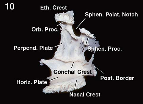
Fig. 2.10 The nasal surface of the palatine bone. The palatine bone is located at the posterior part of the nasal cavity, between the maxilla and the pterygoid process of the sphenoid. It presents horizontal and perpendicular plates. The nasal surface of the perpendicular plate presents two crests: the upper, ethmoidal crest, gives insertion to the middle nasal concha of the ethmoid, and the conchal crest gives insertion to the posterior part of the inferior nasal concha. The superior border of the perpendicular plate presents two processes, orbital and sphenoidal, separated by a deep furrow, the sphenopalatine notch. The orbital process encloses an air sinus. It has three articular and two nonarticular surfaces. It connects anteriorly with the maxilla, posteriorly with the sphenoid concha, and medially with the ethmoid. The superior non-articular surface is part of the floor of the orbit, whereas the lateral non-articular surface faces the pterygopalatine fossa. The sphenoid process is directed medially. Its superior surface connects with the sphenoid concha and the root of the medial pterygoid plate. The medial border of the sphenoid process articulates with the ala of vomer. The sphenopalatine notch is converted into a foramen by the apposition of the sphenoid bone. The foramen links the nasal cavity to the pterygopalatine fossa and transmits the sphenopalatine vessels and posterior superior nasal nerves. The posterior border of the perpendicular plate is continuous above with the sphenoid process, and articulates with the medial pterygoid plate. Eth.: ethmoid, ethmoidal; Horiz.: horizontal; Orb.: orbital; Palat.: palatine; Perpend.: perpendicular; Post.: posterior; Proc.: process; Sphen.: sphenoidal.
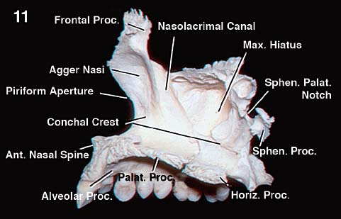
Fig. 2.11 The maxilla and palatine bone have been combined. The nasal surface of the horizontal plate of the palatine bone is concave from side to side and forms the posterior part of the floor of the nasal cavity. Its medial border forms the nasal crest for articulation with the vomer and is continuous with the nasal crest of the maxilla. The anterior border of the perpendicular plate of the palatine bone overlaps the posterior edge of the maxillary hiatus. Both conchal crests of the maxilla anteriorly and of the palatine posteriorly give attachment to the inferior nasal concha. Ant.: anterior; Horiz.: horizontal; Max.: maxillary; Palat.: palatine; Proc.: process; Sphen.: sphenoidal.
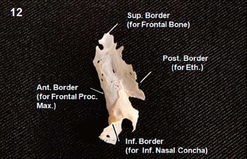
Fig. 2.12 The nasal surface of the lacrimal bone forms part of the middle meatus of the nose. Its upper and posterior part articulate with the ethmoid bone and complete some of the anterior ethmoidal cells. The anterior border articulates with the frontal process of the maxilla. Ant.: anterior; Eth.: ethmoid, ethmoidal; Inf.: inferior; Max.: maxillary; Post.: posterior; Proc.: process; Sup.: superior.
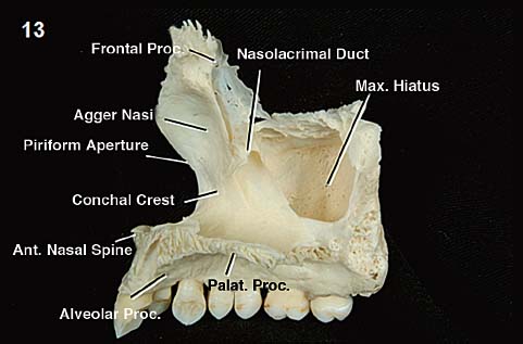
Fig. 2.13 The maxilla and lacrimal bone have been combined. The maxillary contribution to the nasolacrimal canal represents two thirds of its total circumference; the remaining one third is formed by the descending part of the lacrimal bone and lacrimal process of the inferior nasal concha. The nasolacrimal duct opens along the inferior nasal meatus. Ant.: anterior; Max.: maxillary; Palat.: palatine; Proc.: process.
The anterior segment of the floor of the nasal cavity is formed by the maxilla and the posterior quarter by the horizontal plate of the palatine bone (Fig. 2.11).
The upper part of the nasal cavity is called the subfrontal space, olfactory cleft, or nasal carina,1 and understanding its anatomy is important for approaching the anterior nasal fossa, through either the transcribriform or transplanum approaches. The subfrontal space has anterior, middle, and posterior parts, named, respectively, the nasal, ethmoidal, and sphenoidal segments (Figs. 2.21, 2.22, 2.23, and 2.24). The height of the nasal cavity increases from the piriform aperture to the posterior part of the nasal segment. The height is largest at the ethmoidal segment and decreases from its posterior limit to the choana. The nasal spine of the frontal bone and the nasal bones form the roof of the nasal part. The average distance between the posterior wall of the frontal sinus and the anterior edge of the cribriform plate in adults is 12.7 mm.1 The ethmoidal segment presents the ethmoidal and cribroethmoidal foramina. The average length of the cribriform plate on the endocranial side is 20.8 mm, whereas its exocranial surface, facing the nasal cavity, averages 24.7 mm.1 The sphenoethmoidal recess is on the posterior segment. The average length of the planum sphenoidale up to the tuberculum sellae is 20.9 mm.1
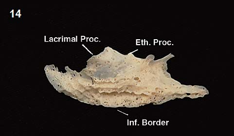
Fig. 2.14 The medial surface of the inferior nasal concha. This surface is convex and presents numerous perforations and grooves for vessels. Its superior border may be divided into three parts. The anterior part articulates with the conchal crest of the maxilla; the posterior part articulates with the conchal crest of the palatine bone; and the middle part presents three processes: lacrimal, ethmoidal, and maxillary. The lacrimal process articulates with the descending part of the lacrimal bone and edges of the nasolacrimal canal of the maxilla, helping the formation of the nasolacrimal duct. The ethmoidal process articulates with the uncinate process of the ethmoid, whereas the maxillary process forms part of the medial wall of the maxillary sinus. Eth.: ethmoid, ethmoidal; Inf.: inferior; Proc.: process.
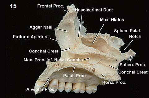
Fig. 2.15 The maxilla, palatine, lacrimal, and inferior nasal concha have been combined. The part of the body of the maxilla below the maxillary hiatus, the maxillary process of the inferior nasal concha, and the part of the perpendicular plate of palatine bone inferior to the conchal crest combine to form the inferior nasal meatus. The opening of the maxillary hiatus is reduced by the apposition of the descending part of the lacrimal concha and the maxillary process of the inferior nasal concha below and the perpendicular plate of palatine behind. Horiz.: horizontal; Inf.: inferior; Max.: maxillary; Palat.: palatine; Proc.: process; Sphen.: sphenoidal.
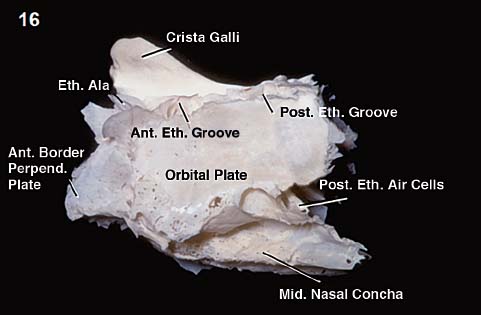
Fig. 2.16 Lateral view of the ethmoid bone. The crista galli is the thick process projecting up from the cribriform plate. Its posterior border gives attachment to the falx. Its anterior border articulates with the frontal bone by the ethmoidal alae. The anterior border of the perpendicular plate articulates with the nasal spine of the frontal bone and the crest of the nasal bones. The orbital plate of the ethmoid is the lateral vertical plate that delimits the ethmoidal air cells, forming the medial wall of orbit and covering the middle and posterior ethmoid air cells. The air cells in front of the orbital plate are completed by the lacrimal bone and the frontal process of the maxilla. The posterior surface of the ethmoidal labyrinths present large, opened air cells to be completed by the sphenoidal concha and the orbital process of the palatine bone. Ant.: anterior; Eth.: ethmoid, ethmoidal; Mid.: middle; Perpend.: perpendicular; Post.: posterior.
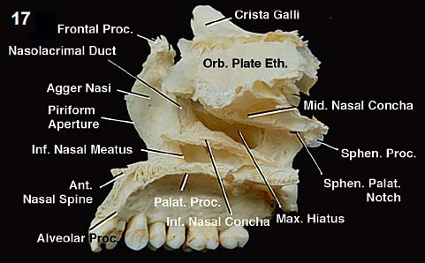
Fig. 2.17 The ethmoid has been added to the bone combination forming the lateral wall of the nasal cavity. It fits along the ethmoidal crest of the perpendicular plate of the palatine, further reducing the maxillary hiatus by the connections of the uncinate process to the maxilla, lacrimal, and inferior nasal concha. Note that the tail of the middle nasal concha is at the level of the sphenopalatine notch. This anatomical fact enables locating the sphenopalatine vessels along the lateral wall of the nasal cavity and planning for mucosal pediculated flaps. Ant.: anterior; Eth.: ethmoid, ethmoidal; Inf.: inferior; Max.: maxillary; Mid.: middle; Orb.: orbital; Palat.: palatine; Proc.: process; Sphen.: sphenoidal.
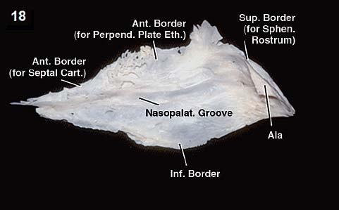
Fig. 2.18 Left lateral view of the vomer. This thin bone forms the posterior-inferior part of the nasal septum. The nasopalatine groove is the deep furrow transmitting the nasopalatine nerve and vessels. The superior border presents a deep furrow bounded by the alae, which connect to the sphenoid conchae, the sphenoidal process of the palatine, and the medial pterygoid plates, forming the superior frame of each choana. Its inferior border attaches to the nasal crest of the maxilla and palatine. The anterior border of the vomer articulates with the perpendicular plate of the ethmoid and the septal cartilage. Ant.: anterior; Cart.: cartilage; Eth.: ethmoid, ethmoidal; Inf.: inferior; Nasopalat.: nasopalatine; Perpend.: perpendicular; Sphen.: sphenoidal; Sup.: superior.
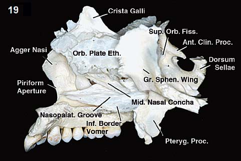
Fig. 2.19 The vomer has been added to its place inferior to the perpendicular plate of ethmoid. It attaches inferiorly to the nasal crest of the maxilla and palatine bones. Posteriorly, the vomer and ethmoid fit along the rostrum of the sphenoid. Ant.: anterior; Clin.: clinoidal; Gr.: greater; Eth.: ethmoid, ethmoidal; Inf.: inferior; Mid.: middle; Nasopalat.: nasopalatine; Orb.: orbital; Proc.: process; Pteryg.: pterygoid; Sphen.: sphenoid; Sup.: superior.
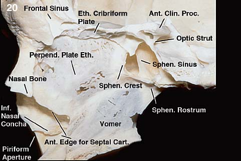
Fig. 2.20 The ethmoidal labyrinth and sphenoid concha have been removed on the left, to expose the bony nasal septum formed by the sphenoid crest, the perpendicular plate of the ethmoid, and the vomer. Ant.: anterior; Cart.: cartilage; Clin.: clinoidal; Eth.: ethmoid, ethmoidal; Inf.: inferior; Perpend.: perpendicular; Proc.: process; Sphen.: sphenoidal.
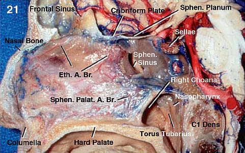
Fig. 2.21 Stepwise dissection of the nasal cavity from the midline to its lateral limit on the right side. The nasal septum has a membranous part at the columella, a cartilaginous part overlying the septal cartilage, and a bony part superoposteriorly. The bones and cartilages of the nasal septum are covered by a layer of periosteum and perichondrium, over which lie the submucosa and the mucosa of the nasal cavity and vestibule. The nasal septum and its mucosal covering vary from 5 to 13 mm in thickness. The supply for the septal mucosa derives mainly from branches of the anterior and posterior ethmoidal arteries and the septal branches of the sphenopalatine artery. A.: artery; Br.: branch, branches; C1: first cervical vertabra; Eth.: ethmoid, ethmoidal; Palat.: palatine; Sphen.: sphenoidal.
Related posts:
 External Versus Endoscopic Approaches for Skull Base Malignancies
 Anatomical Basis of Skull Base Surgery: Skull Osteology


