1 Anatomical Basis of Skull Base Surgery: Skull Osteology
Carolina Martins, Alvaro Campero, Alexandre Yasuda, Luiz Felipe de Alencastro, Shigeyuki Osawa, and Albert L. Rhoton, Jr.
 Introduction
Understanding the osteology of the skull base is a fundamental step in skull base surgery. It enables accurate topographic location and helps tailoring surgical routes to specific skull base areas. This chapter reviews the bony architecture of the anterior, middle, and posterior skull base.
 General Anatomy
The skull is divided into the cranium and facial skeleton. The cranium in its turn is divided into calvaria, which is the domelike superior portion of the cranium, formed by the frontal, parietal, and squamous parts of the occipital and temporal bones and greater sphenoid wings, and the cranial base. The cranial base is formed by the occipital, temporal, ethmoid, and frontal bones arranged around, and connected by, a center element—the sphenoid bone.
The cranial base has an endocranial surface, which faces the brain and is naturally divided into anterior, middle, and posterior fossae (Fig. 1.1) and an exocranial surface (Fig. 1.2), which faces the nasal cavity, sinuses, orbits, pharynx, infratemporal fossae, and pterygopalatine, parapharyngeal, and infrapetrosal spaces.1,2
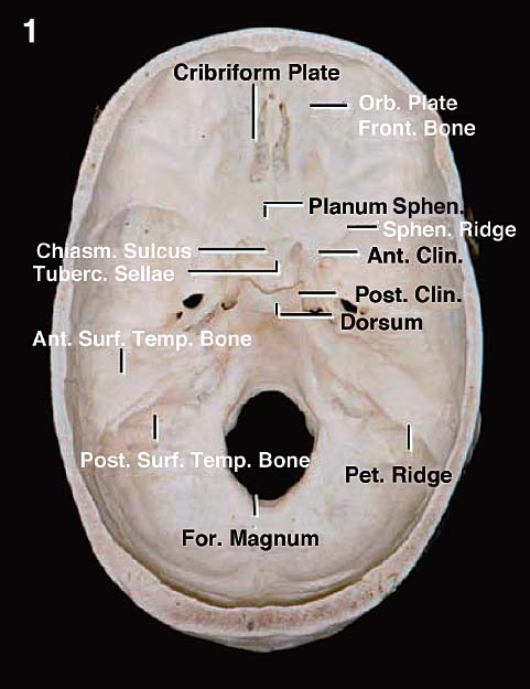
Fig. 1.1 Cranial base: endocranial surface. The upper surface of the anterior cranial base is formed by the frontal bone, which roofs the orbit; the ethmoid bone, which is interposed between the frontal bones and is the site of the cribriform plate; and the lesser wing and the anterior part of the body of the sphenoid, which form the posterior part of the floor of the anterior fossa. The upper surface of the middle cranial base is formed by the greater sphenoid wing and the sphenoid body anteriorly and the upper surface of the temporal bone posteriorly. The posterior part of the cranial base is formed by the temporal and occipital bones. Ant.: anterior; Clin.: clinoid; Chiasm.: chiasmatic; For.: foramen; Front.: frontal; Orb.: orbital; Pet.: petrous; Post.: posterior; Sphen.: sphenoid, sphenoidale; Surf.: surface; Temp.: temporal; Tuberc.: tubercle.
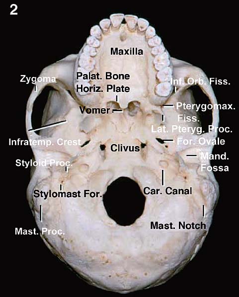
Fig. 1.2 Cranial base: exocranial surface. The exocranial surface is formed mainly by the maxillae, zygomatic, palatine, sphenoid, temporal and occipital bones, and vomer. The maxillae, the orbits, and the nasal cavity are located below the anterior fossa. The anterior part of the hard palate is formed by the maxillae, and the posterior part is formed by the palatine bone. The vomer attaches to the lower part of the body of the sphenoid and forms the posterior part of the nasal septum. The anterior part of the zygomatic arch is formed by the zygoma and the posterior part by the squamosal part of the temporal bones. The mandibular fossa is located below the posterior part of the middle fossa. The infratemporal fossa is located below the greater sphenoid wing and is limited anteriorly by the infratemporal crest. Car.: carotid; Fiss.: fissure; For.: foramen; Horiz.: horizontal; Inf.: inferior; Infratemp.: infratemporal; Lat.: lateral; Mand.: mandibular; Mast.: mastoid; Orb.: orbital; Palat.: palatine; Proc.: process; Pteryg.: pterygoid; Pterygomax.: pterygomaxillary; Stylomast.: stylomastoid.
On the endocranial side of the skull base, the border between the anterior and middle fossa is marked by the sphenoid ridge, joined medially by the chiasmatic sulcus. The border between the middle and posterior fossae is formed by the petrous ridges joined by the dorsum sellae and posterior clinoid processes (Fig. 1.3).
On the exocranial side, the anterior and middle fossae are divided by a transverse line, extending through the pterygomaxillary fissures and pterygopalatine fossae at the upper level, and the posterior edge of the alveolar processes of the maxillae at a lower level. Medially, this corresponds to the attachment of the vomer to the sphenoid bone. The middle and posterior cranial fossae are separated on each side by a transverse line crossing near the posterior border of the vomer-sphenoid junction, foramen lacerum, carotid canal, jugular foramen, styloid process, and mastoid tip (Fig. 1.4).
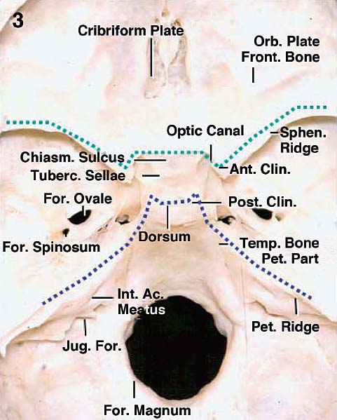
Fig. 1.3 On the endocranial side of the skull base, the border between the anterior and middle fossa is marked by the sphenoid ridge, joined medially by the chiasmatic sulcus (dotted light blue line), and the border between the middle and posterior fossae is formed by the petrous ridges joined by the dorsum sellae and posterior clinoid processes (dotted dark blue line). Ac.: acoustic; Ant.: anterior; Chiasm.: chiasmatic; Clin.: clinoid; For.: foramen; Front.: frontal; Int.: internal; Jug.: jugular; Orb.: orbital; Pet.: petrous; Post.: posterior; Sphen.: sphenoid; Temp.: temporal; Tuberc.: tubercle.
Each of the three skull base areas has a center and two lateral parts. The center parts are arranged as a midline corridor and comprise, on the endocranial side, the cribriform area, planum, sellae, clivus, and craniovertebral junction. On the exocranial side, this center corridor encompasses the nasal cavity, sphenoid sinus, and the pharynx.
In the center corridor, the anterior, middle, and posterior skull base areas are close together and bridged by the body of the sphenoid.
 Anatomy of the Anterior Skull Base
The anterior endocranial surface is formed by the combination of three bones—frontal, ethmoid, and sphenoid (Fig. 1.5). The orbital plates of the frontal bones form most of the lateral parts of this fossa, are the roof of the orbital cavities, and give support to the dura and orbital gyri of the frontal lobe. The medial gap between the orbital plates is filled by the cerebral surface of the ethmoid bone, presenting the crista galli and cribriform plates. The crista galli gives attachment to the falx, whereas the cribriform plates give support to the olfactory bulbs and are traversed by the olfactory fila. Posteriorly, the anterior fossa is closed by the lesser wings of the sphenoid laterally and the sphenoid body medially. In this way, the medial portion of the anterior fossa is formed by three bones, whereas the lateral part, which covers the orbit and optic canals, is formed only by two, the orbital plate of frontal bone and the lesser sphenoid wings, on each side.
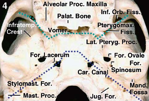
Fig. 1.4 On the exocranial side, the anterior and middle fossae are divided by a transverse line, extending through the pterygomaxillary fissures and pterygopalatine fossae at the upper level, and the posterior edge of the alveolar processes of maxillae at a lower level. Medially, this corresponds to the attachment of vomer to the sphenoid bone (dotted light blue line). The middle and posterior cranial fossae are separated on each side, by a transverse line crossing near the posterior border of vomer–sphenoid junction, foramen lacerum, carotid canal, jugular foramen, styloid process, and mastoid tip (dotted dark blue line). Car.: carotid; Fiss.: fissure; For.: foramen; Inf.: inferior; Infratemp.: infratemporal; Jug.: jugular; Lat.: lateral; Mand.: mandibular; Mast.: mastoid; Orb.: orbital; Palat.: palatine; Proc.: process; Pteryg.: pterygoid; Pterygomax.: pterygomaxillary; Stylomast.: stylomastoid.
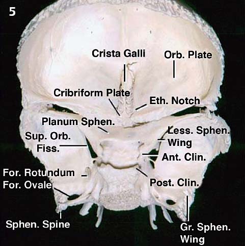
Fig. 1.5 The frontal, ethmoid, and sphenoid bones combine to form the anterior fossa, which is divided into medial and lateral portions. The medial part, covering the upper nasal cavity and sphenoid sinus, is formed by the crista galli and the cribriform plate of the ethmoid bone anteriorly and the planum of the sphenoid body posteriorly. The lateral part, which covers the orbit and the optic canal, is formed by the frontal bone and the lesser wing of the sphenoid bone, which blends medially into the anterior clinoid processes and points toward the middle fossa. Ant.: anterior; Clin.: clinoid; Eth.: ethmoid; Fiss.: fissure; For.: foramen; Gr.: greater; Less.: lesser; Orb.: orbital; Post.: posterior; Sphen.: sphenoid; Sup.: superior.
On the exocranial side, the lateral portion of the anterior skull base is on the top of the orbit and maxillary sinus. Medially, it corresponds to the sphenoid sinus of sphenoid body and the ethmoid sinuses, on top of the nasal cavity (Fig. 1.6). The most posterior portion of the medial exocranial anterior surface is related to the sphenoid, whereas the medial and anterior thirds are related to the ethmoid bone.
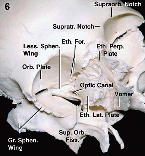
Fig. 1.6 On the exocranial side, the anterior cranial base is divided into a medial part related to the ethmoidal and sphenoidal sinuses and nasal cavity below, and a lateral part that corresponds to the orbit and maxilla. The ethmoid bone forms the anterior and middle thirds of the exocranial surface, and the sphenoid body forms the posterior third of the medial part. The ethmoid presents the perpendicular plate that joins the vomer in forming the nasal septum and two lateral plates located in the medial wall of the orbits. The lateral plates separate the lateral wall of the nasal cavity and the orbit. The main foramina of the region are the anterior and posterior ethmoidal foramina located in the superomedial orbital wall, along the frontoethmoidal suture, which transmit the ethmoidal nerves and arteries; the supraorbital and supratrochlear notches or foramina, transmitting the arteries and nerves of the same name; and the optic canal, which transmits the optic nerve and ophthalmic artery. The superior orbital fissure is located between the lesser and greater sphenoidal wings on the lateral side of the optic canal. It transmits the oculomotor, trochlear, ophthalmic, and abducens nerves, a recurrent meningeal artery, and the superior and inferior ophthalmic veins. Eth.: ethmoid; Fiss.: fissure; For.: foramen; Gr.: greater; Lat.: lateral; Less.: lesser; Orb.: orbital; Perp.: perpendicular; Sphen.: sphenoid; Sup.: superior; Supraorb.: supraorbital; Supratr.: supratrochlear.
The bony nasal septum, which is formed by the vomer and perpendicular plate of the ethmoid and attached to the sphenoid crest and rostrum, divides the nasal cavity along the midline, whereas the lateral plates of the ethmoid bones separate the nasal cavity from each orbit (Figs. 1.7 and 1.8).
Some foramina and grooves connect the endocranial and exocranial surfaces and transmit vascular and neural structures in this area. The foramen cecum in the midline serves as the site of passage of an emissary vein; the cribriform plate is pierced by the filaments of the olfactory nerve; the supraorbital grooves, on the superior orbital limits, are related to the frontal branch of the first trigeminal division; the anterior and posterior ethmoidal canals, located along the suture line formed by the frontal and ethmoid bones, transmit the anterior and posterior ethmoidal nerves and arteries; the superior orbital fissure, located between the lesser and greater sphenoidal wings, transmit the superior ophthalmic vein and the first division of the trigeminal, oculomotor, trochlear, and abducens nerves; and the optic canals between the anterior and posterior roots of the anterior clinoid processes transmit the optic nerve and the ophthalmic artery.
 Anatomy of the Middle Skull Base
The endocranial surface of the middle fossa is formed by the sphenoid and temporal bones. The division between these bones usually is not easy to see unless one is focusing on the sphenoid spine, the most posterior prominence of the sphenoid bone, just posterolateral to the foramen spinosum. From this point it is possible to follow the sphenopetrosal and sphenosquamosal sutures (Fig. 1.9). The middle cranial base has medial and lateral parts. The medial part is formed by the body of the sphenoid, whereas the lateral parts result from the combination of lesser and greater sphenoid wings and squamous and petrous parts of the temporal bone. The medial portion of the middle cranial base is the sellae, whereas the most lateral portions are the temporal fossae. Between these two areas, on each side, are the parasellar regions. The parasellar regions are probably the smallest areas of the skull base with the highest concentration of important neural and vascular structures, as they house the cavernous sinuses.
The sphenoid contributes to the middle fossa mainly with its body, the greater and lesser wings. Laterally, the lesser sphenoid wings form the sphenoid ridges. Medially, the lesser wings are connected to the sphenoid body through the anterior root, and they form the roof of the optic canal and are continuous with the sphenoid planum. At the center of the planum is the sphenoid jugum, a faint ridge, which is the remnant of the fusion of the ossification centers. The posterior root of the anterior clinoid process, also called the optic strut, separates the optic canals above from the superior orbital fissure below. The chiasmatic sulcus is located posterior to the planum. On each side of the chiasmatic sulcus are the endocranial openings of the optic canals. Posteriorly, the chiasmatic sulcus is separated from the sellar cavity by the tuberculum sellae. The posterior limit of the sellae is composed of the dorsum and posterior clinoid processes, which are the medial boundaries between the middle and posterior cranial fossae (Fig. 1.10).
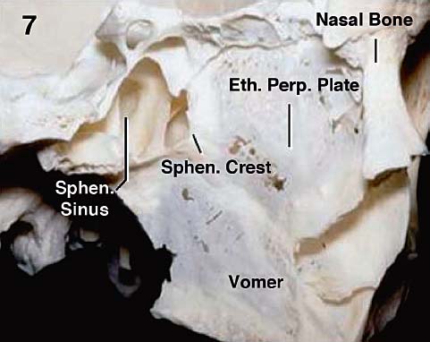
Fig. 1.7 The osseous nasal septum is formed by the attachment of the perpendicular plate of the ethmoid and vomer at the sphenoidal crest. Eth.: ethmoid; Perp.: perpendicular; Sphen.: sphenoid.
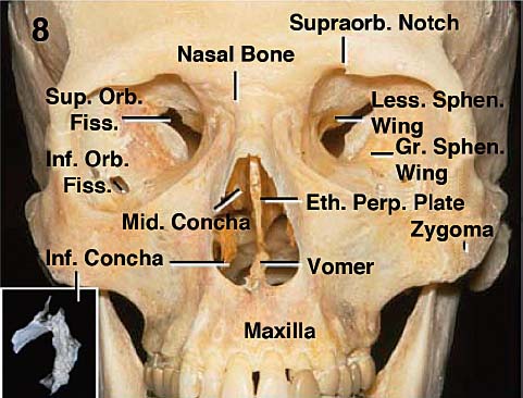
Fig. 1.8 Anterior norma. The orbital rim is formed by the frontal bone, zygoma, and maxilla. The nasal bone is interposed above the anterior nasal aperture, between the maxillae. The nasal cavity is located between the ethmoid bone above and the maxillae, palatine bones, and pterygoid processes of the sphenoid below. It is roofed by the frontal and ethmoid bones, and the floor is formed by the maxillae and palatine bones. The nasal septum forms the medial wall of the nasal cavities. The nasal conchae are located on the lateral walls of the nasal cavity. The inferior concha (inset) is a separate bone, and the middle and superior concha are appendages of the ethmoid bone. Eth.: ethmoid; Fiss.: fissure; Gr.: greater; Inf.: inferior; Less.: lesser; Mid.: middle; Orb.: orbital; Perp.: perpendicular; Sphen.: sphenoid; Sup.: superior; Supraorb.: supraorbital.
Related posts:
 Anesthesia in Endoscopic Skull Base and Brain Surgery
The Endonasal Transplanum-Transtuberculum Endoscopic Approach in Pituitary Adenomas
Endoscopic Transnasal Craniectomy and the Resection of Extensive Craniopharyngiomas
External Versus Endoscopic Approaches for Skull Base Malignancies
Combined Cranio-Endoscopic Approach
Anatomy of the Orbit and Related Structures
Anesthesia in Endoscopic Skull Base and Brain Surgery
The Endonasal Transplanum-Transtuberculum Endoscopic Approach in Pituitary Adenomas
Endoscopic Transnasal Craniectomy and the Resection of Extensive Craniopharyngiomas
External Versus Endoscopic Approaches for Skull Base Malignancies
Combined Cranio-Endoscopic Approach
Anatomy of the Orbit and Related Structures
Stay updated, free articles. Join our Telegram channel

Full access? Get Clinical Tree


