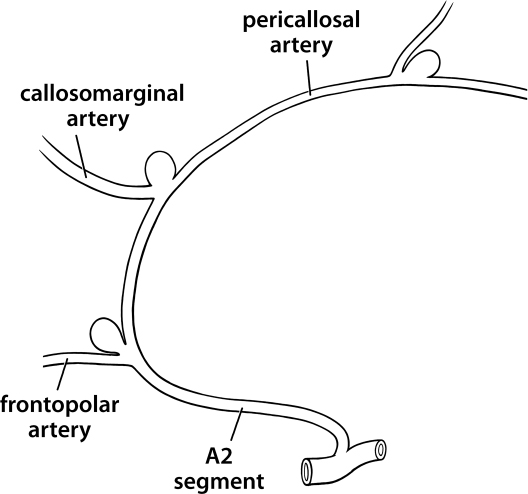
8 These lesions—defined as aneurysms arising distal to the anterior communicating complex—are relatively uncommon and are frequently overlooked on initial diagnostic angiography (especially when occurring as unruptured companions to ruptured aneurysms). They are very common in the setting of multiple aneurysms and occur with disproportionate frequency in series of mycotic aneurysms. Their unique location makes them somewhat awkward to expose, in part because of the unfamiliarity of the approach, and in part because, as a rule, proximal control can only be achieved late in dissection. Unlike aneurysms at more common locations, the major morbidity of surgery for distal anterior cerebral artery (DACA) aneurysms relates neither to the risk of early aneurysmal rupture nor ischemia to the distributions of the afferent arteries, but rather to injury, either by direct retraction or by venous infarction, involving the cortex adjacent to the route of surgical exposure. For that reason, whenever feasible we prefer a nondominant, parasagittal exposure. The normal anatomy of the DACA involves a variable number of proximal branches to the mesial frontal cortex, the largest of which is referred to as the orbital-frontal artery (Fig. 8.1). These branches always arise inferior to the genu of the corpus callosum and may infrequently be the site of DACA aneurysms. Fig. 8.1 Common locations for DACA aneurysms. The most proximal large branch of the DACA is the frontopolar artery that routinely arises inferior to or at the level of the genu and projects anteriorly along the mesial frontal cortex in the general direction of the frontal pole. This origin is the second most common site of DACA aneurysms. The largest branch of the DACA is usually the callosomarginal artery, which may arise inferior, anterior, or dorsal to the genu of the corpus callosum. This artery and its three terminal branches supply the majority of the mesial frontal cortex and anastomose with anterior and superior branches of the middle cerebral artery in the “watershed” area of the parasagittal cortex. As might be anticipated because of its size, the origin of the callosomarginal artery is the most frequent site of DACA aneurysms. The anterior cerebral artery (ACA) continues after the takeoff of the callosomarginal artery as the pericallosal artery (A4 segment). This vessel runs in the cistern of the corpus callosum for a variable distance prior to terminating in several smaller cortical branches. In its distal segment it commonly anastomoses with the posterior pericallosal artery, a branch of the posterior cerebral artery. Aneurysms along the A4 segment are uncommon, but when they do occur, they are frequently mycotic in etiology. Variations in the anatomy of the DACA are very common, with azygous A2 segments; proximal DACA bifurcations giving rise to long, parallel, callosomarginal and pericallosal arteries; and numerous small callosomarginal branches from a large parent DACA being perhaps the most frequent.
Aneurysms of the Distal Anterior Cerebral Artery
General
Anatomy
Vascular
Cerebral
Related posts:
 The Evolution of the Intracranial Aneurysm Surgeon
Operating Room Considerations: Equipment, Setup, and Culture
The Evolution of the Intracranial Aneurysm Surgeon
Operating Room Considerations: Equipment, Setup, and Culture
 Surgical Management of Previously Coiled Aneurysms
 Aneurysms of the Internal Carotid Artery at the Origins of the Posterior Communicating and Anterior Choroidal Arteries
 Aneurysms of the Proximal Basilar Artery
 Aneurysms of the Vertebral Artery at the Origin of the Posterior-Inferior Cerebellar Artery
Stay updated, free articles. Join our Telegram channel

Full access? Get Clinical Tree


