7 Anomalous Innervations
Martin–gruber Anastomosis
Routine Ulnar Conduction Study: Pseudo-Conduction Block between the Wrist and Below-Elbow Sites
The MGA may be recognized during routine ulnar motor studies, recording the abductor digiti minimi, stimulating at the wrist and below-elbow sites (Figure 7–1). If the anastomotic fibers innervate the abductor digiti minimi, a characteristic pattern results: a drop in the ulnar compound muscle action potential (CMAP) amplitude is seen between the wrist and the below-elbow stimulation sites (Figure 7–2). With stimulation at the wrist, the CMAP reflects all motor fibers innervating the hypothenar muscles, including those that have crossed over more proximally from the median nerve. Stimulation at the below-elbow site activates fewer fibers, however, as this stimulation site is above the cross-over. Thus, the portion of fibers innervating the abductor digiti minimi that originate from the median nerve have already crossed over in the forearm and, therefore, do not contribute to the CMAP. The differential diagnosis of this pattern (i.e., higher amplitude distally than proximally) includes the following:
• Excessive stimulation of the ulnar nerve at the wrist resulting in co-stimulation of the median nerve
• Submaximal stimulation of the ulnar nerve at the below-elbow site
• Conduction block of the ulnar nerve between the wrist and below-elbow sites
• An MGA with crossing fibers innervating the hypothenar muscles
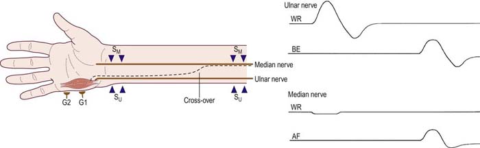
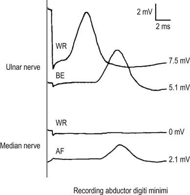
Whenever there is a >10% drop in amplitude between the wrist and below-elbow sites on routine ulnar motor studies, median nerve stimulation should always be performed at the wrist and at the antecubital fossa while recording the hypothenar muscles to check for an MGA. If no MGA is present, a small positive deflection usually is recorded with both the wrist and antecubital fossa stimulation sites, reflecting a volume-conducted potential from median muscles (see Chapter 2). If an MGA is present, a small positive volume-conducted potential will be present with median nerve stimulation at the wrist; however, median stimulation at the antecubital fossa will evoke a small CMAP over the abductor digiti minimi. The amplitude of the CMAP evoked by stimulating the median nerve at the antecubital fossa (recording the hypothenar muscles) will approximately equal the difference between the CMAP amplitudes evoked with ulnar nerve stimulation at the wrist and below-elbow sites (recording the hypothenar muscles). However, it is important not to overstimulate the median nerve at the antecubital fossa, resulting in co-stimulation of the ulnar nerve at the elbow, and thereby giving the appearance of an MGA when none truly exists. This can be avoided by slowly moving the stimulator from the median nerve at the antecubital fossa to the ulnar nerve at the elbow, stimulating at several points. In a true MGA, the CMAP that is evoked with median nerve stimulation at the antecubital fossa will briefly disappear as the stimulator is moved toward, but not over, the ulnar nerve at the elbow. It will then reappear with ulnar nerve stimulation at the elbow.
Ulnar Conduction Study: Proximal Martin–Gruber Anastomosis and Pseudo-Conduction Block between the Below-Elbow and Above-Elbow Sites
In patients with ulnar neuropathy at the elbow, one of the classic electrophysiologic findings is conduction block across the elbow, whereby a drop in the CMAP amplitude is seen between the below-elbow and above-elbow sites during routine ulnar motor studies (see Chapter 19). This pattern is not typically confused with an MGA, because the drop in CMAP amplitude in a typical MGA occurs between the wrist and below-elbow sites, mimicking a conduction block in the forearm, not across the elbow. However, very rarely, the cross-over fibers of the MGA are very proximal, and if stimulated, will contribute to the CMAP amplitude at the below-elbow site. In contrast stimulation above the elbow will not excite these cross-over fibers, thereby giving the impression of a conduction block across the elbow. It is in these cases, where the below-elbow stimulation might occur below the MGA, that an MGA may result in the mistaken diagnosis of ulnar neuropathy with conduction block at the elbow (Figure 7–3). This is especially apt to occur when the below-elbow stimulation site is too distal, making it more likely that the below-elbow stimulation occurs below the MGA, thereby exciting the cross-over fibers.
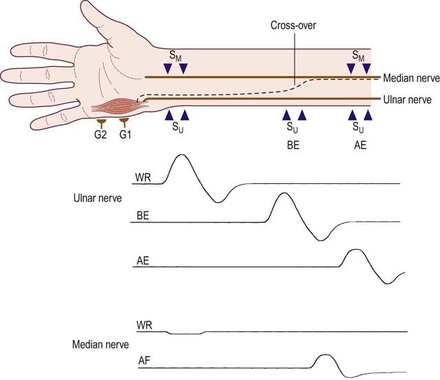
In one anatomic study of cadavers found to have an MGA, the anastomosis joined the ulnar nerve an average of 8.4 cm (range 5–12) distal to the medial epicondyle, whereas electrophysiologic studies have suggested the possibility of an MGA even more proximal, as close as 3 cm distal to the medial epicondyle. Thus, if the below-elbow site is stimulated 3 cm or further distal to the medial epicondyle (especially >5 cm), there is a possible risk of a proximal MGA mimicking the pattern of an ulnar neuropathy with conduction block at the elbow. As ulnar neuropathies across the elbow typically occur either at the elbow or at the cubital tunnel (under the aponeurosis of the flexor carpi ulnaris), the below-elbow stimulation site needs to be at least 2 cm distal to the medial epicondyle, which is the most distal location of the cubital tunnel. This underscores the need to stimulate the below-elbow site of the ulnar nerve at the proper location, 3 cm distal to the medial epicondyle (see Chapter 10), and not more distally. In addition, one should always look for an MGA in any patient with a conduction block of the ulnar nerve at the elbow without any other supporting abnormalities to suggest an ulnar neuropathy at the elbow.
Ulnar Conduction Study Recording the First Dorsal Interosseous: Pseudo-Conduction Block between the Wrist and Below-Elbow Sites
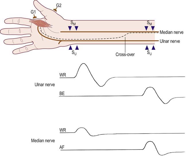
The most common MGA (Figure 7–4) occurs with crossing over of median-to-ulnar fibers supplying the FDI. Nevertheless, this anastomosis is not often recognized during routine ulnar motor nerve conduction studies because the abductor digiti minimi is the muscle most often recorded for routine ulnar motor studies. However, it is not an uncommon finding when ulnar motor studies are performed recording the FDI. The FDI is commonly recorded in two situations: (1) looking for a lesion of the deep palmar motor branch of the ulnar nerve (i.e., ulnar neuropathy at the wrist), and (2) when evaluating a suspected ulnar neuropathy at the elbow (see Chapter 19).

Stay updated, free articles. Join our Telegram channel

Full access? Get Clinical Tree


