10 Routine Upper Extremity, Facial, and Phrenic Nerve Conduction Techniques
Median Motor Study (Figure 10–1)

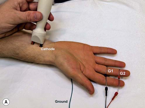
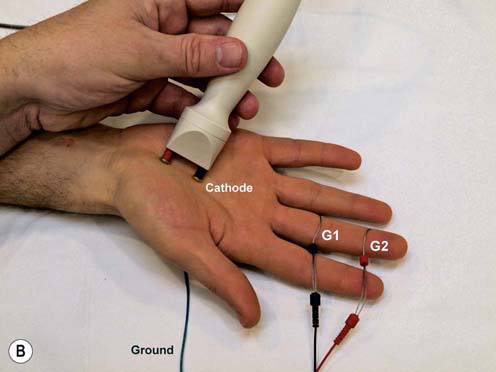
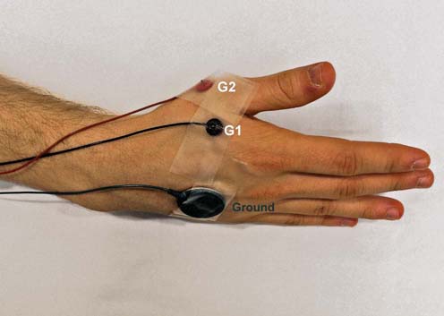
Median Motor Palmar Study (Figure 10–2)
Key Points:
• The APB is innervated via the recurrent thenar motor branch of the median nerve, which runs into the palm and then curves back to the thenar muscles.
• A palm/wrist CMAP amplitude ratio >1.2 implies some conduction block across the wrist.
• Calculation of conduction velocity is not reliable because of the short distances and the course of the recurrent branch of the thenar motor branch.
• If palm stimulation results in baseline distortion due to stimulus artifact, the anode should be rotated until a suitable baseline is obtained.
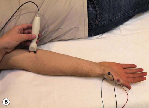
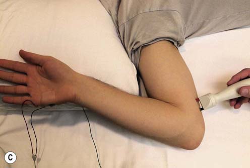
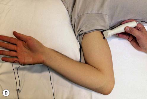
Median Sensory Study (Figure 10–3)
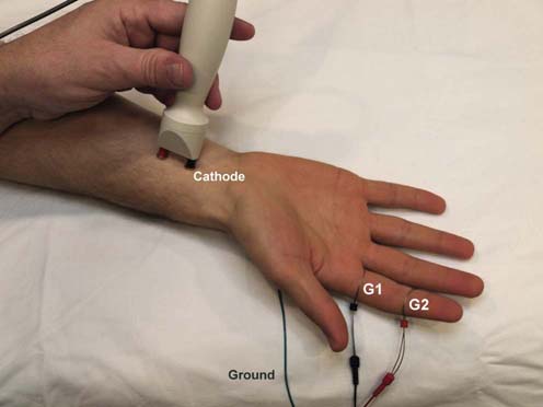
Key Points:
• The study is easy to perform.
• Antidromic study described. For orthodromic study, recording and stimulation sites are reversed.
• A volume-conducted motor potential occasionally may obscure the sensory potential in antidromic studies. If this occurs, have the patient slightly spread their fingers and stimulate again.
• Stimulation also can be performed proximally at the antecubital fossa, similar to the median motor study; however, the proximal sensory response is normally smaller and more difficult to record because of normal temporal dispersion and phase cancellation.
• Digits 1 and 4 both are partially innervated by the median nerve and can also be used for median sensory studies.
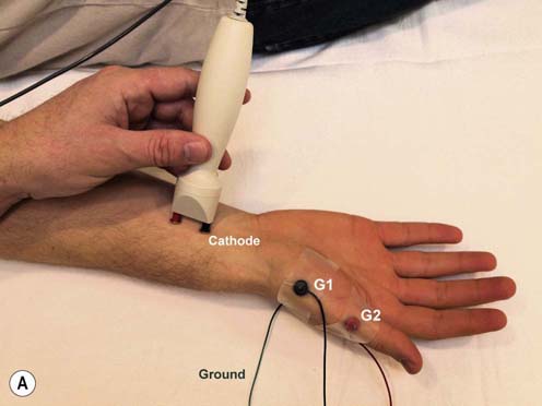
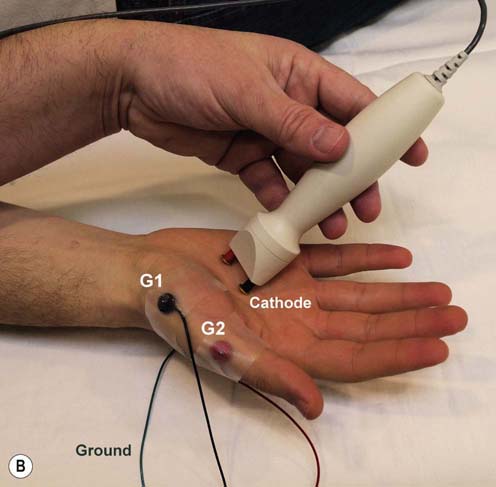
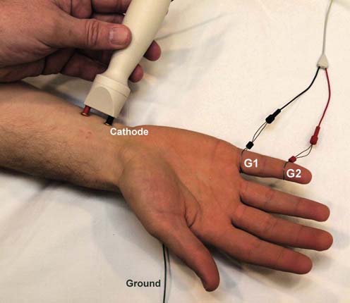
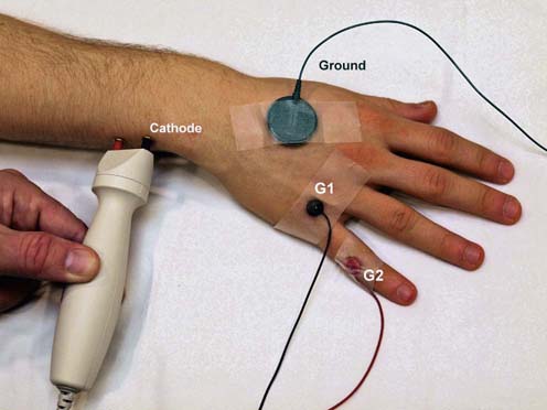
Median Sensory Palmar Study (Figure 10–4)
Key Points:
• A palm/wrist sensory nerve action potential (SNAP) amplitude ratio >1.6 implies some conduction block across the wrist.
• It is essential to obtain a clear onset latency at both sites (electronic averaging is often helpful).
• At the palm stimulation, stimulus artifact may contaminate the onset latency. It is essential to obtain a clear onset latency at both the palm and wrist sites. If palm stimulation results in baseline distortion due to stimulus artifact, the anode should be rotated until a suitable baseline is obtained.
• From this study, the conduction velocities for the wrist-to-digit 3 segment and the palm-to-digit 3 segment are displayed on the machine. On some EMG machines, the wrist-to-palm segment conduction velocity is also calculated and displayed on the machine. However, if the EMG machine does not calculate the conduction velocity, it must be mathematically calculated, by subtracting the palm-to-digit 3 onset latency from the wrist-to-digit 3 onset latency. Then a conduction velocity for the wrist–palm segment (i.e., across the carpal tunnel) can be calculated by taking the distance (7 cm) and dividing it by the calculated latency. The wrist-to-palm conduction velocity (i.e., across the carpal tunnel) is normally faster than the palm-to-digit 3 segment. In carpal tunnel syndrome, there is a reversal of this pattern, with relative slowing of the wrist-to-palm segment (see Chapter 17).
• Note that any distance can be used at the wrist and at the palm. However, if the palm-to-digit 3 distance is half the distance of the wrist-to-digit 3, the mathematical calculation is much simpler (see Chapter 17).
• This study is also known as the median segmental sensory study, as two sensory segments of the median nerve (wrist-to-palm and palm-to-digit) are compared.
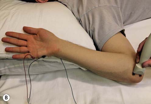
Ulnar Motor Study (Figure 10–5)
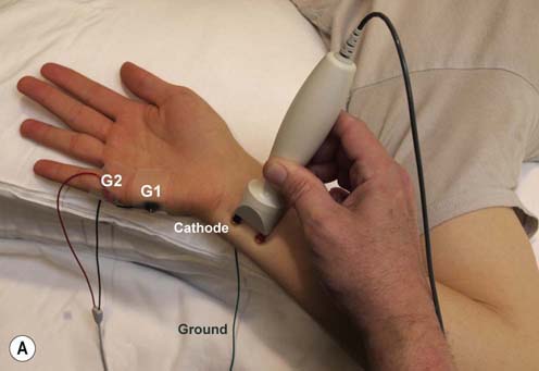
Stimulation Sites:
Wrist: Medial wrist, adjacent to the flexor carpi ulnaris tendon
Below elbow: 3 cm distal to the medial epicondyle
Above elbow: Over the medial humerus, between the biceps and triceps muscles, at a distance of 10–12 cm from the below-elbow site
Axilla (optional): In the proximal axilla, medial to the biceps over the axillary pulse
Key Points:
• The optimal position is with the elbow flexed between 90° and 135°. If performed in a straight-elbow position, factitious slowing across the elbow will be seen due to underestimation of the true nerve length.
• Higher current intensity usually is needed to achieve supramaximal stimulation at the below-elbow site compared with the wrist and above-elbow sites because the nerve lies deep to the flexor carpi ulnaris muscle at this location.
• Stimulation must be at least 3 cm distal to the medial epicondyle at the below-elbow site to ensure that stimulation is distal to the cubital tunnel, a common site of ulnar nerve compression at the elbow. However, if stimulation at the below-elbow site is too distal (>4 cm), the nerve is very deep and very difficult to stimulate, reinforcing that the optimal stimulation site is 3 cm distal to the medial epicondyle.
• Always perform wrist, below-elbow, and above-elbow stimulations. If only the wrist and above-elbow stimulations are performed, one can miss ulnar slowing across the elbow.
• The distance across the elbow must be measured along a curved line, with the elbow flexed, and not as a straight line. This approximates the true anatomic course of the nerve.
• If the CMAP amplitude at the below-elbow site is more than 10% smaller than that at the wrist, consider a Martin–Gruber anastomosis.
Ulnar Sensory Study (Figure 10–6)
Key Points:
• Antidromic study described. For orthodromic study, stimulation and recording sites are reversed.
• A volume-conducted motor potential occasionally may obscure the sensory potential in antidromic studies. If this occurs, have the patients slightly spread their fingers and stimulate again.
• May be abnormal in ulnar neuropathy or lower trunk brachial plexopathy (e.g., thoracic outlet syndrome).
• Stimulation also can be performed proximally at the below- and above-elbow sites, similar to the ulnar motor study; however, the proximal sensory responses are normally smaller and more difficult to record because of normal temporal dispersion and phase cancellation.
Dorsal Ulnar Cutaneous Sensory Study (Figure 10–7)
Key Points:
• Supramaximal stimulation usually can be achieved with low stimulation intensities (e.g., 5–15 mA) because the nerve is quite superficial.
• Often helpful to compare side-to-side amplitudes in cases where one side is symptomatic and the other is not.
• Always spared in lesions of the ulnar nerve at Guyon’s canal.
• May be abnormal in some, but not all, cases of ulnar neuropathy at the elbow.
Deep Ulnar Motor Branch Study (Figure 10–8)
Recording Site:
Key Points:
• The deep ulnar motor branch often is preferentially affected in lesions of the ulnar nerve at Guyon’s canal.
• Recording the FDI may be more useful than recording the ADM for demonstrating focal slowing of the ulnar nerve across the elbow.
• G2 must be on the metacarpal–phalangeal joint of the thumb; if G2 is place on the metacarpal–phalangeal joint of the index finger, there will always be an initial positive deflection of the CMAP.
• Always perform the wrist, below-elbow and above-elbow stimulations. If only the wrist and above-elbow stimulations are performed, one can miss ulnar slowing across the elbow.
• Stimulation must be at least 3 cm distal to the medial epicondyle at the below-elbow site to ensure that stimulation is distal to the cubital tunnel, a common site of ulnar nerve compression at the elbow. However, if stimulation at the below-elbow site is too distal (>4 cm), the nerve is very deep and very difficult to stimulate, reinforcing that the optimal stimulation site is 3 cm distal to the medial epicondyle.
• If the CMAP amplitude at the below-elbow site is more than 10% smaller than that at the wrist, consider a Martin–Gruber anastomosis.

Stay updated, free articles. Join our Telegram channel

Full access? Get Clinical Tree


