Superficial landmarks include:
• hyoid C3
• thyroid cartilage C4–5
• cricoid C6
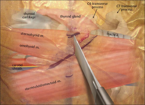
• A horizontal incision is made just medial to the sternocleidomastoid muscle (SCM).
• A decision on a right- or left-side approach should be made based upon surgeon comfort.
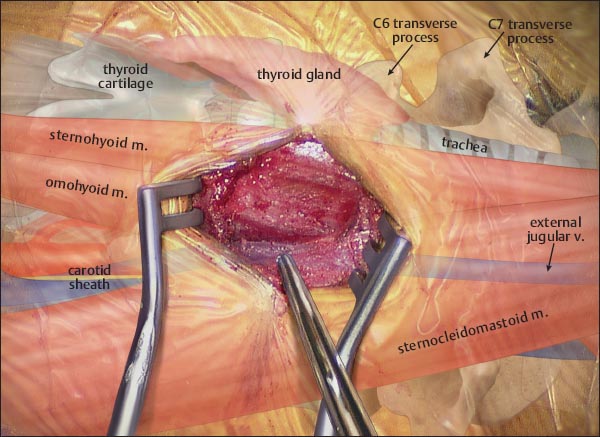
• The platysma is divided in line with the skin incision.
• The external jugular vein helps to identify the tracheoesophageal groove.
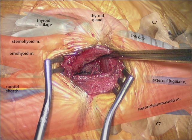
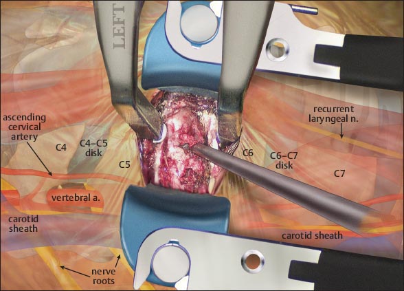
• The SCM and carotid sheath are retracted laterally.
– The tracheoesophageal complex is retracted medially.
– The recurrent laryngeal nerve lies in the tracheoesophageal groove.
– The cartoid sheath contains:
 the internal jugular vein
 the cartoid artery
 the vagus nerve
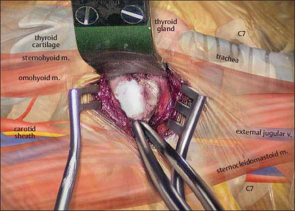
• The longus colli are swept laterally, exposing the superficial disk space.
– The sympathetic chain lies superficial to the longus colli; therefore, retractors should be placed deep into this muscle.
• A knife or electrocautery device can be used to perform the annulotomy.
Stay updated, free articles. Join our Telegram channel

Full access? Get Clinical Tree


